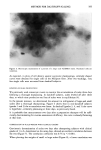
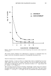
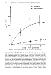
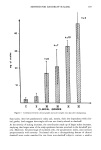
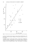
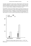
j. Soc. Cosmet. Chem., 39, 179-190 (May/June 88) A new method to quantify scaling in dandruff D. SAINT-LEGER, A. M. KLIGMAN, and T. J. STOUDEMAYER, Laboratoires de Recherche de l'Oreal, Dgpartement de Biophysique, 93601 Aulnay-Sous-Bois, France (D.S.-L. ), Department of Dermatology, University of Pennsylvania, Philadelphia, PA (A.M.K.), and Biosearch, Inc., 3408-50 B Street, Philadelphia, PA 19134 (T.J.S. ). Received February 1 O, 1988. Synopsis Previous methods for appraising scaling in dandruff lack accuracy and hence reproducibility in different laboratories. We have developed a gravimetric procedure which directly determines the quantity of scales and also permits separate appraisal of small and large scales. The scalp is shampooed in a standard way with lauryl ether sulfate. The suspended scales are first filtered through a mesh screen of pore size 200 Ix, trapping large scales. The tiltrate is then passed through a screen enabling the collection of small scales. The second tiltrate contains single cells which selectively can be retained in an 8-Ix Millipore filter, stained, and counted. The small and large scales are dried and weighed. Additional information can be obtained by disaggre- gating the scales in a sonicator, after which the percentage of nucleated cells can be assessed. The latter value, the parakeratotic index, is a measure of the degree of inflammation. We found that (1) single cells comprise the majority of the corneocytes shed in dandruff (2) the percentage of nucleated cells is far greater on large, compared to small scales, confirming the underlying inflammatory process in dandruff, a feature not clinically evident (3) the quantity of scales and their distribution corre- lates well with clinical grades but is more reliable and (4) an appreciable quantity of scales can be consis- tently collected from the scalps of persons who do not have dandruff. The method can be applied to any scaling disease of the scalp. INTRODUCTION Dandruff is a non-inflammatory scaling disorder of the scalp. This is a clinical defini- tion requiring the observer to exclude other scaly scalp diseases such as psoriasis, atopic dermatitis, tinea capiris, etc. Whether dandruff is simply low-grade seborrheic derma- titis is in dispute but is not germane to our purpose since the method we shall describe is equally applicable to that condition. The experienced technician can grade the severity of dandruff by visual estimation of the quantity of scales. This may be adequate for assessing the efficacy of anti-dandruff 179

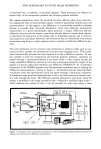
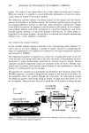
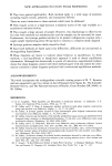
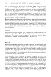
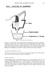
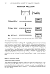
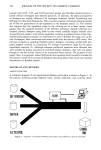
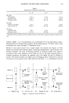
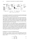
180 JOURNAL OF THE SOCIETY OF COSMETIC CHEMISTS treatments but is not sufficiently sensitive to differentiate among agents which may bring dandruff under control but which differ significantly in speed of response, degree of response, and relapse time after treatment is stopped. Naked-eye inspection is semi- quantitative at best and has the limitation that clinical severity is determined not only by the quantity of scales but by their size as well. The presence of some large flakes will lead to higher gradings, whereas dominantly small flakes will be underestimated. Scales are visible aggregates of coherent corneocytes they vary considerably in size, and the difference between small and large flakes may be thousands of cells. Then, too, the mere dispersal of aggregates by surfactants could substantially lower the dandruff grade without influencing the process which characterizes dandruff, namely, an excessive pro- duction of horny cells by a hyperproliferative epidermis. Various methods have been devised to overcome the subjective limitations of clinical grading (1-3). Vacuuming the scalp has been tried but is unreliable because it removes mainly the larger scales, leaving an unknown amount behind the same applies to combing techniques. Counting the corneocytes obtained by scrubbing a small site with a detergent (4) has been more successful as a quantitative method but it, too, has important handicaps. The correlation with clinical grades is weak because only single cells are counted in the hemocytometer aggregates are overlooked. A novel method, recently published in the Japanese literature (5), utilizes an infrared light source and a photodetector to scan dandruff scales as they are sucked through a cylinder. The drop in voltage is a function of the volume of each scale, the number and size of which has been computer-calculated by a formula. This complex instrument is not destined for routine use and its accuracy has still to be assessed. We shall now describe a method for quantifying scales these are defined as the smallest aggregate of corneocytes that can be seen with the naked eye. MATERIALS AND METHODS The subjects were young adult males with varying degrees of dandruff, excluding sebor- rheic dermatitis. A trained technician rated the severity of dandruff on a 0 to 10 scale, as previously described (6). Grade 3 corresponds to marginal dandruff these subjects generally do not seek treatment. Grade 5 is moderate dandruff and 6 is rather intense scaling. The great majority of subjects fell within the grade range of 3 to 6. Anti-dan- druff treatments were stopped for three weeks prior to the study. PROCEDURE With the subject lying down, the scalp was washed in a standard way for 45 seconds by a single technician using 5 ml of a 6.25% aqueous solution of sodium lauryl ether sulfate (WITCO). The scalp was then rinsed clean of scales with 2 to 4 liters of fresh water as needed, carried out over a sink with an arrangement for collecting the wash water in a large plastic bucket (Figure 1). This was repeated once more and the washings pooled. Thus there were two successive 5-ml shampooings. As summarized in Figure 2, the washings were poured into a funnel
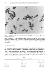
Purchased for the exclusive use of nofirst nolast (unknown)
From: SCC Media Library & Resource Center (library.scconline.org)