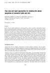
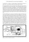
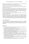
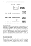
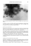
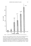
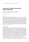
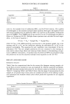
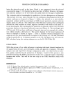
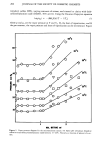
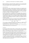
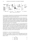
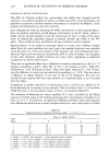
NEW APPROACHES TO STUDY PHASE PROPERTIES 163 a horizontal line, or isotherm, in the phase diagram. These techniques are referred to, respectively, as the temperature gradient and lyotrope gradient methods. The sample preparations create the potential for more efficient phase data collection, but making the most of this potential requires a means of rapid phase identification and characterization. In this regard, x-ray diffraction is a particularly powerful technique because it provides direct structural information. Since each molecular arrangement characteristic of a given mesomorphic phase generates a unique diffraction pattern, diffraction data provide the means to positively identify phases in single phase regions, to determine the proportion of each phase type in multiphase regions, and to structur- ally characterize each phase. However, the low photon flux of conventional x-ray sources necessitates long (hours to days) exposure times to produce satisfactory diffraction pat- terns. This time limitation can be overcome with synchrotron x-radiation when used in con- junction with a suitable two-dimensional live-time x-ray imaging device. This combi- nation dramatically decreases the time required to record a diffraction pattern. To ana- lyze samples in both the temperature and lyotrope gradient methods, capillaries are passed through a synchrotron-derived x-ray beam while a video camera records the image-intensified diffraction pattern in live-time continuously along the length of the sample in a process called time-resolved x-ray diffraction (TRXRD) (4-6). A schematic illustration of the TRXRD apparatus and the associated equipment used in making the diffraction measurements is shown in Figure 4. Synchrotron-derived monochromatic x-radiation enters the experimental hutch and passes through a delimiting collimator. A translation stage positioned perpendicular to the x-ray beam moves the sample capil- lary tube through the beam. The diffraction pattern generated at each point along the sample is amplified in the image intensifier tube--basically a modified night scope-- which enhances the pattern so that it can be recorded on video tape using a video Figure 4. Schematic of the experimental arrangement at the Cornell High Energy Synchrotron Source for making time-resolved x-ray diffraction measurements.

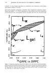
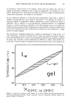
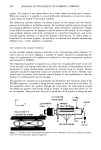
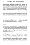
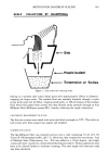
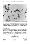
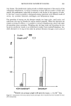
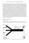
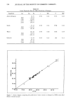
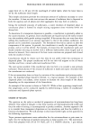
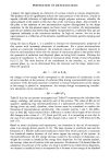
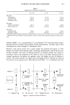
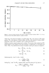
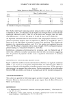
164 JOURNAL OF THE SOCIETY OF COSMETIC CHEMISTS camera. The output of the camera feeds into a video cassette recorder and a monitor. With this system it is possible to record diffraction information in live-time contin- uously along the length of the sample capillary. The diffraction patterns indicate the phases present in the sample and the relative amount of each phase in multiphase regions. By recording capillary position along with the changing diffraction pattern on video tape, phase boundaries, signified by changes in the diffraction pattern, are assigned to precise points in the sample. In the tempera- tures gradient method, each point corresponds to a specified temperature, and in the lyotrope gradient method, to a particular lyotrope concentration. To collect phase in- formation for the entire diagram, the procedure is repeated with samples representing different lines--either isopleths or isotherms. THE TEMPERATURE GRADIENT METHOD In this method, samples represent isopleths in the corresponding phase diagram. To collect data for an entire diagram, a number of sample capillaries encompassing the range of compositions to be analyzed are subjected to the same temperature gradient and analyzed by TRXRD. The temperature gradient is supported on a metal rod. A regulatable heater at one end of the rod and a circulating water bath at the other end create a linear gradient which is monitored by using thermocouples positioned at intervals along its length. Sample capillaries are placed lengthwise around the perimeter of the rod with a thermal com- pound used to promote good thermal contact (Figure 5) and equilibrated so that the gradient is communicated into the samples. The gradient rod, mounted on a goniometer and attached to the translation stage of the TRXRD apparatus, is rotated to bring the first sample in line with the x-ray beam. As the translation moves the capillary through the x-ray beam, the video system records the diffraction pattern continuously along its length. A single scan takes about 20-30 sec to complete. After each scan, the rod is rotated like a Colt revolver to bring the next circulating x-ray l y water batIn beam X gonlometer sample • head 'x• 11aries. heater motorized X-Y translation stage ! gradient rod thermacouples variac Figure 5. Schematic of the experimental arrangement used in making live-time x-ray diffraction measure- ments with the temperature gradient apparatus.
Purchased for the exclusive use of nofirst nolast (unknown)
From: SCC Media Library & Resource Center (library.scconline.org)