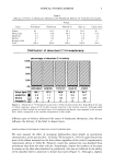
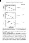
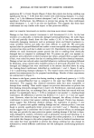
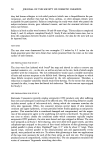
14 JOURNAL OF THE SOCIETY OF COSMETIC CHEMISTS •/-hydroxamate, dimethyl casein (DMC), ot-keto glutaric acid, NADH, NAD, cadav- erine dihydrochloride, histamine hydrochloride, and putrescine dihydrochloride. Tri- ethanolamine hydrochloride, Tris base, dithiothreitol (DTT), calcium chloride dihy- drate, and disodium EDTA were Fisher Biotech TM grade. [ 1,4-•4C]-putrescine dihydro- chloride (115 mCi/mmol) was purchased from Amersham. HAIR SAMPLES AND PREPARATION The human hair used in all phases of this work was obtained from several individual donors with known hair-treatment histories. All hair was chemically unprocessed, com- monly referred to as "virgin" hair, having received only normal shampooing, condi- tioning, and styling products and procedures. Hair used with the optical assay was obtained from an adult female, cut approximately 2 cm from the scalp at the back of the neck, and used in 0.5 cm lengths. The hair used for the first through third isotope experiments was obtained from a child of about eight years of age. This hair was removed from a ponytail cut about 20 cm below the scalp. The fourth isotope experi- ment used three different sources of hair obtained from adult female donors. This hair was collected by donors on a day-to-day basis as it exfoliated during grooming. For isotope experiments, only sections falling within 15 cm of the "root" end were used. The child's hair, even though originally sampled farther from the scalp, was probably the least damaged, due to infrequent grooming. In any event, all hair used in this work had a high probability of having minimal cuticle damage from weathering and other cuticle degradation processes. All hair samples were washed before use with a 30% solution of triethanolamine lauryl sulfate/sodium laureth sulfate (TEALS/SLES). This consisted of a 1-2 minute wash by hand followed by a 5-minute rinse in distilled water, except where noted below. A more rigorous procedure was adopted for the isotope experiments. At this point, the hair was processed as bundles of ten fibers, glued together at their root ends. Hair bundles were wet with water and wound onto stainless steel hair clips, shown in Figure 1. Clips were grouped together and subjected to collective washing and rinsing steps prior to incu- bation. Loaded clips were soaked for 10-30 minutes in a stirred beaker of 5% TEALS/ SLES and _rinsed under-flowing distilled water for a half hour. OPTICAL ASSAY A coupled enzyme assay for monitoring transglutaminase activity was adapted from an enzymatic assay for ammonia (37). The concentration of glutamate dehydrogenase was increased to improve assay response to low levels of ammonia. The assay solution (150 mM triethanolamine hydrochloride buffer, 12 mM ot-keto glutarate, 0.6 mM ADP, 5 mM CaCI2, 5 mM DTT, 2.5 mg/ml dimethyl casein (DMC), pH 7.8) was sterile- filtered through a 0.45-•tm filter and stored frozen. DMC, an amino-blocked glu- tamine-containing protein substrate, was omitted from incubations with hair. NADH solution (16.67 mg/ml NADH in water, stabilized with NaHCO3) was kept at 4øC and prepared fresh daily. Stock solutions of amines were buffered to normality with HC1 or prepared directly as amine hydrochloride salts. The glutamate dehydrogenase solution was used as purchased from Sigma in 50% glycerol (1200 U/ml). The transglutaminase stock solution was approximately 1.0 unit/ml in 10 mM Tris buffer, pH = 7.5,

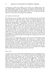
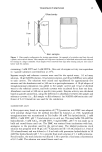
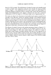
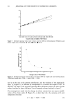
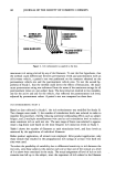
TRANSGLUTAMINASE 15 Glued ends ß •Hai r sample Figure 1. Hair sample configuration for isotope experiments. An example of a stainless steel hair clip and a plastic microvial are shown. Hair samples and clips were incubated in individual microvials and removed for rinsing in a larger container. Hair samples were removed from clips after rinsing and put into scintil- lation vials for analysis. containing 2 mM DTT and 2 mM EDTA. One unit of enzyme activity was equivalent to 1 •mole substrate converted/min at 25øC. Separate sample and reference cuvettes were used for the optical assay. 1.0 ml assay solution, 10 •1 NADH solution, 10 •1 amine solution, and 40 •i GiDHase were added to each cuvette. The solutions were mixed and equilibrated for approximately five minutes and the absorbance read at 340 nm in a spectrophotometer. A 10-•1 aliquot of transglutaminase preparation was added to the sample cuvette and 10 •i of distilled water to the reference cuvette, and both cuvettes were incubated for at least one hour. Absorbance was read at 340 nm at specific time points. Enzyme activity was calculated based on •moles amine/min, using the difference in absorbance between the sample and reference cuvettes (i.e., AA sample vs AA reference). An NADH mM extinction coef- ficient of 6.22 i/mmol/cm was used for the calculation. RADIOISOTOPE ASSAY A filter paper assay based on incorporation of [14C]-putrescine into DMC was adapted with minimal change from the method described by Lorand et al. (38). Lyophilized transglutaminase was reconstituted in Tris buffer (10 mM Tris hydrochloride, 2 mM EDTA, 2 mM DTT, pH 7.5) and stored on ice until use. The assay buffer (50 mM Tris hydrochloride, 5 mM CaCi 2, 20 mM DTT, 5 mg/ml DMC, pH 7.5) was made up in bulk and stored frozen until use. The DMC was omitted from incubations with hair, analogous to the procedure used for the optical assay. The labeled putrescine stock solution was prepared with 50 •Ci of [14C]-putrescine (@ 115 mCi/mmol in 1.0 ml of 2% ethanol/water) and was diluted to 2.0 ml with cold putrescine hydrochloride in 50 mM Tris hydrochloride (pH 7.5) to provide final stock solution of 1.2 mM putrescine. For all studies, the stock solution was diluted 1:10 to provide a final assay concentration of 0.12 mM. The solution was stored frozen until use.
Purchased for the exclusive use of nofirst nolast (unknown)
From: SCC Media Library & Resource Center (library.scconline.org)