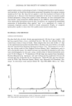
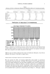
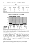
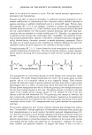
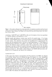
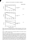
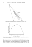
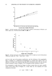
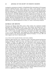
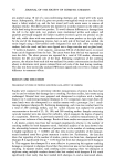
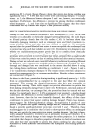
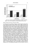
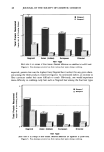
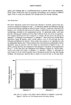
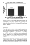
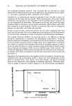
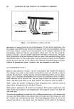
2 JOURNAL OF THE SOCIETY OF COSMETIC CHEMISTS applied hydrocarbons vs physiological lipids. Utilizing both fluorescent and ultrastruc- tural methods, we found that hydrocarbons penetrated throughout the stratum corneum interstices (6,7) but that they did not appear to enter the nucleated cell layers of the epidermis (6,7). Because of the theoretical importance of the issue of cutaneous and/or internal malignancy arising from uptake of these materials, we have investigated this issue further, using isotopically labeled alkanes of two different chain lengths to quan- titate hydrocarbon movement across the skin. In addition, we compared the transdermal penetration of hydrocarbons when applied "neat" or as ingredients of a variety of commonly employed cosmetic vehicles of mixed composition. Finally, we compared hydrocarbon uptake in intact skin with uptake across acetone-treated skin. MATERIALS AND METHODS ANIMALS AND MATERIALS Pig skin (back skin of adult, female pigs approximately 130 days of age weight = 100 kg) was acquired from a local, commercial butcher. The skin was obtained fresh, and stored frozen within 6 hours of death. These samples then were used within 2-7 days in the experimental procedures described below. Hairless male mice (hr/hr), 6-8 weeks old were acquired from Simonsen Laboratories (Gilroy, CA). 3H-docosane (53.97 mCi/ mg) was custom made by New England Nuclear (Boston, MA). Immediately prior to use in the pig skin assays, the labeled docosane was repurified by preparative radio-thin- layer chromatography on silica gel, using a mobile phase of hexane:diethylether:acetic acid (70:30:2) (Figure 1). •4C-hexadecane (250 •tCi/mg) was acquired from Amersham- Buchler (Braunschweig, Germany), and used without further purification because the material had been prepurified by the manufacturer. TESA •53205 tape was manufac- tured by TESA Tape Industries (Sparta, Maine), resp. Beiersdorf AG (Hamburg, Ger- many). An electrolytic water analyzer (Model W, Type SPR-DMU Meeco, Inc., War- 489 1 3 7 [r=] Figure 1. Thin-layer chromatographic profile of 3H-docosane utilized for the absorption studies in porcine skin. Only material migrating between 6 and 8.5 cm was employed.

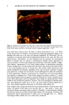
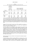
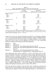
TOPICAL HYDROCARBONS 3 rington, PA) was used to measure transepidermal water loss (TEWL) (8). Chloral hydrate (Rugby Laboratories, Inc., Rockville Center, NY) was utilized for anesthesia of mice by intraperitoneal injection (0.4 mg/g body weight). Acetone, propylene glycol, petroleum ether, and Scintisafe 30% scintillation cocktail were purchased from Fisher Scientific (Pittsburgh, PA). Vaseline © petroleum jelly was from Chesebrough-Ponds (Trumbull, CT). Petrolatum "WITCO," polydecene "Nexbase 2006 FG," mineral oil "USP," soya oil, and w/o cream were laboratory samples from Beiersdorf AG. Liquid scintillation counting was performed on liquid scintillation spectrometers (models LS3801 and LS1800 Beckman Instruments, Houston, TX). Hionic Fluor scintillation cocktail and Solvene 350 tissue solubilizer were acquired from Packard Instruments (Downers Grove, IL). FLUORESCENCE MICROSCOPY Skin biopsies of petrolatum-treated areas from hairless mice were collected, snap-frozen in liquid nitrogen, sectioned at 4 •m, and stained with nile red to show the distribution of topical "neat" petrolatum 2-4 hours after application to full-thickness skin (7). All frozen sections were viewed in a Leitz Ortholux II microscope equipped for epifluores- cence (Leica, Inc., Deerfield, IL). IN VITRO MODEL Pig skin was prepared for penetration studies by scraping away the subdermal fat, gently dry-shaving the outer stratum corneum surface, cutting the skin into 5-cm-diameter discs (thickness •3-4 ram), and freezing at -70øC until needed. Immediately prior to percutaneous transport experiments, the skin discs were thawed, mounted in glass penetration cells (Frantz design, custom made in Hamburg, Germany) at 32øC (skin temperature), and filled with 0.9 M sodium chloride with 1% bovine serum albumen. Test mixtures of petrolatum "WITCO," polydecene "Nexbase 2006FG," mineral oil "USP," soya oil, and a bland cosmetic w/o cream were combined with either 40 ng 3H-docosane in 3 mg of the test mixture/cm 2, or 200 ng •4C-hexadecane in 5 mg of the test mixture/cm 2. Mixtures were applied topically to intact skin discs, incubated for 24 hours, and then harvested as the following skin fractions for liquid scintillation count- ing: a) Skin surface--gently scraped with a spatula and/or three-fold wiping with cotton wool b) Stratum corneum--about 15-20 strippings with "TESA" tape until reaching a "shiny" surface, indicative of removal of the bulk of the stratum corneum c) Epidermal nucleated layers--full-thickness skin discs applied epidermis-side-downward on an 80øC hot plate for 45 seconds, followed by separation of epidermis and dermis with forceps, after prior tape stripping. d) Dermis--the application area (25-mm diameter) cut out of heat-split, whole dermis [see (c) above] e) Receptor fluid--two aliquots taken at the end of each 24-hour incubation. Receptor fluid, surface swabs, and tape strippings with attached stratum corneum were placed in Hionic Fluor and counted directly. Epidermis and dermis samples first were
Purchased for the exclusive use of nofirst nolast (unknown)
From: SCC Media Library & Resource Center (library.scconline.org)