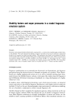
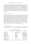
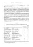
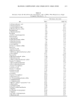
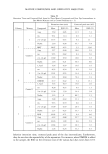
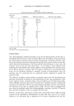
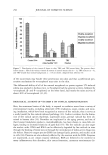
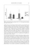
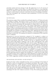
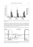
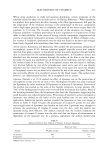
268 JOURNAL OF COSMETIC SCIENCE meation and bioconversion of a provitamin to vitamins C and E in hairless mouse skin. Infinite dosing was used for the study. It was found that the rate of appearance of vitamin E after bioconversion increased gradually during the 72-h study period. Because vitamin E is a very lipophilic compound, a considerable amount that was bioconverted in the viable skin was expected to diffuse back into the stratum comeurn until the concentra- tion in the stratum comeurn reached a steady state. This process was believed to be very time-consuming. However, the provitamin was not completely bioconverted to vitamins C and E in the hairless mouse skin. The yield of bioconversion was calculated to be about 96%. The authors have also cautioned that the bioconversion is dependent and influ- enced by the activity and distribution of enzymes in the skin therefore, the findings obtained in the hairless mouse skin may not be directly applicable to humans. However, the authors concluded that such bioconversion of the provitamin to vitamins E and C simultaneously would occur in human skin because of its enzyme esterase distribution. It can be stated of this experiment that the authors did not take any special precautions to maintain enzyme viability during the 72 h. Norkus et •L (104) demonstrated using an i, vitro study that topical application of ot-tocopheryl acetate gel to hairless mouse skin results in a significant increase in free ot-tocopherol levels. This bioconversion was further enhanced by exposure to UVB irradiation. They demonstrated that topical application of 5% ot-tocopheryl acetate gel to hairless mouse skin resulted in significant accumulation of ot-tocopheryl acetate in skin tissue. The ot-tocopheryl acetate is absorbed and retained by skin tissue following topical application. Skin from placebo gel-treated animals either exposed or not exposed to UVB irradiation (groups A and B) contained low levels of free ot-tocopherol. Skin ot-tocopherol levels were significantly increased (P 0.001) following topical applica- tions of ot-tocopheryl acetate (groups C and D). In addition, skin levels of free ot-to- copherol are significantly greater (P 0.001) in UVB-irradiated animals compared to non-UVB-irradiated animals that received identical daily topical ot-tocopheryl acetate treatments. The results are summarized in Table III. This study, however, does not allow ot-tocopherol mobilized from other tissue sources to be distinguished from that obtained by the hydrolysis of topically applied ot-tocopheryl acetate. Trevithick and Mitton (105) studied the bioconversion of ot-tocopheryl acetate to free ot-tocopherol across mouse skin in vivo. Radiolabeled ot-tocopheryl acetate was not only well absorbed but also transported laterally in the skin after absorption. ot-Tocopheryl Table III c•-Tocopherol in Mouse Skin From Four Different Treatment Groups Group Vitamin E acetate UVB exposure o•-Tocopherol in skin (pg/g) A - - 2.8 _+ 0.54 • B - + 4.15 _+ 0.86 • C + - 19.91 + 2.68 b D + + 34.01 _+ 14.38 Results are mean _+ SD. • ANOVA comparisons found statistically significant between the indicated groups and groups C or D (P 0.001). b ANOVA comparisons found statistically significant between the indicated groups and group D (P 0.001). Adapted from reference 104.

SKIN DELIVERY OF VITAMIN E 269 acetate was hydrolyzed to free tx-tocopherol approximately 4.5-5 % of the tx-tocopheryl acetate was hydrolyzed in the skin within 24 hours to free tx-tocopherol. The lipophilic tx-tocopheryl acetate was thought to be absorbed by diffusion through the cell mem- brane to the cytoplasm within individual cells, where it may be hydrolyzed by intra- cellular esterases and/or lipases. Esterases are also thought to occur in sweat, and vitamin E acetate could undergo extracellular hydrolysis before its absorption by the skin in such species. Kramer-Stickland and Liebier (106) used a sensitive gas-chromatography-mass- spectrometry assay to distinguish between endogenous tx-tocopherol and deuterium- labeled tx-tocopherol resulting from hydrolysis of topically applied deuterated tx-tocoph- eryl acetate on mouse skin. This permitted an unambiguous characterization of the contribution of topically applied o•-tocopheryl acetate to total epidermal o•-tocoperhol content. There was a modest hydrolysis of the deuterated ester prodrug to the deuterated tocopherol in unirradiated animals, reflecting a tenfold increase in total epidermal o•-tocopherol. Pretreatment of mice with UVB caused an even greater degree of hydrol- ysis. If the skin was pretreated with tx-tocopherol before UVB exposure, the UVB enhancement of o•-tocopheryl acetate hydrolysis was abolished, indicating that UVB was the trigger for increased hydrolysis. The acetate at a 5.3 pmol (50 mg of a 5% cream) dose did not completely protect the skin from photodamage. Hydrolysis in the unirra- diated mice seemed to be dependent on the amount of the acetate absorbed. Hydrolysis was saturable over a 24-h period, and increases in the acetate dose did not result in a corresponding increase in its hydrolysis. Hydrolysis in irradiated mice was time- dependent but not limited by absorption. tx-Tocopheryl acetate was hydrolyzed to the active antioxidant tx-tocopherol in mouse skin, and hydrolysis and the esterase activity was potentiated by UVB irradiation. Gensler and co-workers (107) reported the inability of mouse skin to cleave tx-tocopheryl acetate and succinate to the antioxidant form tx-tocopherol and their consequent inabil- ity to prevent photocarcinogenesis. The authors tested the capacity of the thermostable forms of tx-tocopherol, its acetate and succinate esters, to prevent UV-induced skin cancer in mice in vivo. At 17 weeks after treatment, skin was removed and concentrations of o•-tocopherol and its esters were detected chromatographically. The authors found that most of the vitamin E applied to skin in the ester form remained unchanged, with limited cleavage to free tx-tocopherol. There was only 10-30% as much tx-tocopherol as there was o•-tocopheryl acetate or succinate (wt/wt), respectively. Also, the chronic topical administration of the ester forms was unable to prevent UV-induced photocar- cinogenesis. The authors have reasoned that the limited capacity of mouse skin to cleave the ester forms to the antioxidant form of o•-tocopherol may explain the inability of o•-tocopheryl acetate or succinate to prevent photocarcinogenesis. IN VITRO AND IN VIVO STUDY In vitro study. Increasing evidence supports the contention that in vitro permeability studies can somewhat accurately predict in vivo absorption and possibly cutaneous me- tabolism. This is only true if studies take into account potential problem areas. During such in vitro experiments it is important to ensure that skin viability is maintained throughout the time duration of the experiment. Cadaver skin should be used cautiously the permeability should be checked with standard compounds. Wester et al. (108) have
Purchased for the exclusive use of nofirst nolast (unknown)
From: SCC Media Library & Resource Center (library.scconline.org)