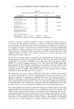
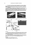
150 JOURNAL OF COSMETIC SCIENCE also been shown to increase heat shock protein expression in mammalian cells (12,13). In cell culture systems, 1,25-dihydroxyvitamin D3 has been reported to increase kera tinocyte differentiation (14,15). 1,25 Dihydroxyvitamin D3 has also been shown to increase p53 and c-fos expression in keratinocytes (16). Kitano et al. (17) observed a decrease in keratinocyte cell growth with 1,25-dihydroxyvitamin D3. However, others have reported that 1,25-dihydroxyvitamin D3 increased cell growth in normal keratinocyte proliferation (18). This contradiction may be due to the biphasic dose response effects of 1,25-dihydroxyvitamin D3 on keratinocyte proliferation (19). These results suggest that 1,25-dihydroxyvitamin D3, and possibly its precursor 7-de hydrocholesterol, may induce a stress response in keratinocytes. The present investiga tion was undertaken to determine if 7-dehydrocholesterol induces heat shock proteins and if this induction results in any clinical benefit when 7-dehydrocholesterol (7-DHC) is applied topically to human skin. MATERIALS AND METHODS Normal human epidermal keratinocytes (NHEK Clonetics) were grown in KBM media (CloneticsY supplemented with hydrocortisone (5 x 10- 4 mg/ml), epidermal growth factor (1 x 10- 7 mg/ml), insulin (5 x 10- 3 mg/ml), bovine pituitary extract (2 ml), and GA-1000 (antibiotic) (0.5 ml). NHEKs, approximately 80% confluent, were treated with 10- 6 M 7-dihydrocholesterol. Normal human epidermal keratinocytes (NHEK) were grown in media supplemented with human keratinocyte growth serum (HKGS). Total RNA was recovered with TRizol Reagent (Life Technologies) and treated with Dnase to remove any trace DNA. Total RNA levels were quantitated by fluorescent staining with RiboGreen (Molecular Probes) and by gel electrophoresis on 1 % agarose. The cDNA was prepared with the RETROscript kit (Ambion). The external standard was cyclophilin (Ambion) and the internal standard was G6PDH. cDNA was heated for ten minutes prior to amplification and was amplified with T aq polymerase (DNA Master SYBR Green I, Roche Molecular Biochemicals) in 20-µl reactions containing the fol lowing amounts of cDNA as template: 1 µg for cyclophilin, 0.5 µg for G6PDH, and 5 µg for HSP70A, HSP90 alpha, HSP90 beta, and HSP27. PROTEIN LEVELS OF HSP70 NHEK were grown to 75% confluency in six-well plates before being treated with different doses of 7-DHC (in EtOH). These treatments were carried out for 24 hours. Following the treatment, the keratinocytes were harvested by trypsination and centrifu gation. The cells were then resuspended in lysing buffer (150 mmol/1 NaCl, 50 mmol/1 Tris buffer pH 8.0, 1 % NP-40) and sonicated for three one-minute intervals with a cone attachment on a model W-225 sonicator (Heat Systems-Ultrasonics, Inc., Farmingdale, NY). The HSP70 ELISA kit from StressGen (Victoria, Canada) was used to quantify the levels of HSP70 in the NHEK supernatants. Hyperosmotic stress with sorbitol was used as a positive control to demonstrate that heat shock protein is induced in these cells (33).

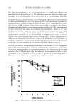
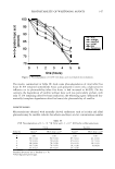
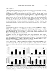
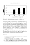
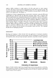
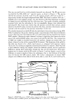
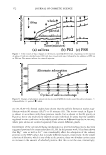
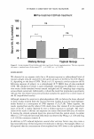
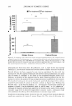
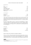
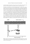
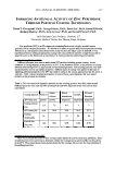
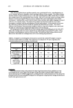
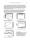
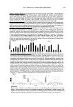
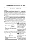
NHEK AND INCREASED HSPs 151 CLINICAL RESULTS 7 Dehydrocholesterol was applied to the skin in ethanol at concentrations of 20 µg/cm2, 40 µg/cm2, and 60 µg/cm2. Healthy volunteers, ages 18-50, males and females with Fitzpatrick type I-II skin, participated in the study. The skin on the backs of the panelists was evenly colored and free of blemishes and stretch marks. The subjects were free of dermatologic or ophthalmologic problems. The source of radiation was the Berger solar simulator. The test material was patched on the backs of the panelists and allowed to absorb for two hours for the induction of HSPs. After two hours, the patch was removed and allowed to air dry for 30 minutes. The MED of the panelists was obtained on the treated site as well as on an adjacent untreated site. The same procedure was repeated with the vehicle (alcohol), which is also suspected to induce heat shock proteins. RESULTS A heat shock of 42 degrees for one hour was utilized to increase the mRNA for HSP 27, HSP90 alpha, HSP90 beta, and HSP70A in normal human keratinocytes in culture. Treating normal human keratinocytes with 7-DHC (10- 6 M) for 24 hours was also effective at inducing the mRNA for HSP 27, HSP90 alpha, HSP90 beta, and HSP70A (Figure 1) as compared to the untreated cells. The ability of 7-DHC to induce the protein levels of the major heat shock protein, HSP70, was investigated. 7-DHC was found to significantly increase the levels of HSP70 protein levels in normal human keratinocytes (Figure 2). This increase was found to be dose-dependent. At 1.25 µM, 2.5 µM, and 10 µM (10- 5 M), there were 27%, 66%, and 71 % increases of HSP70 in keratinocytes, respectively. Topical application of 7-DHC to human skin provided a slight protection from UVB induced erythema (Figure 3). This suppressive effect on UVB erythema was dose dependent. Alcohol was used as the vehicle and did not provide any protection against UVB. 60000 -�-------------ll-····· ............ , i 50000 1 .I 40000 � 30000 i 20000 ii 10000 � untreated . e j1 i untreated heat shock 0.1 uM 7-DHC 1 uM 7-DHC 120000 j 100000 . 80000 . ,, 60000 � 40000 .2: 20000 � 0 untreated heat shock 0.1 uM 7-DHC 1 uM 7-DHC 1 Figure 1. Expression of mRNA for heat shock proteins in normal human keratinocytes. Keratinocytes were treated for 24 hours with 1.0 and 0.1 µM of 7-DHC. Messenger RNA was isolated and probed by RT-PCR for HSP27 (A), HSP90alpha (B), HSP90 beta (C), and HSP70A (D). All points are the mean ± SE. *p 0.05 vs untreated keratinocytes.
Purchased for the exclusive use of nofirst nolast (unknown)
From: SCC Media Library & Resource Center (library.scconline.org)