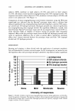
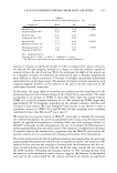
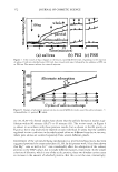
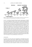
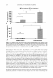
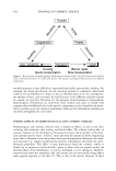
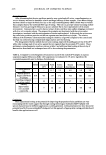
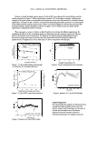
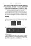
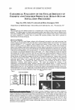
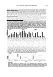
2003 ANNUAL SCIENTIFIC MEETING 215 THE FIBRONECTIN NETWORK DURING AGING: A MISSING CELL CONNECTIVITY Eric Perrier, F. Pivard, S. Grenier and V. Andre Coletica, F-69007, Lyon, France The connective tissue of the skin has been extensively studied but there is still main information missing. The main scaffolds, namely the collagen and the elastic networks, are now more or less elucidated as far as their three-dimensional organization and functions are concerned. For instance, collagens molecules are able to be assembled into collagen fibrils and then fibers that are responsible for mechanical properties of the skin. On the other hand, the complex elastic fibers network is also tremendously important for the plasticity of the skin, and plays a pivotal role in the intrinsic and UV-related aging phenomena. A third network has been poorly studied until now but is also essential to the connective tissue organization as well as the cell-cell and cell-matrix interactions: the fibronectin network. Fibronectin designates a family of glycoproteins (about twenty members) synthesized by alternative splicing. Produced in insoluble form by the connective tissue's fibroblasts, it is found within cells at the cell surface and on extra-cellular levels. Fibronectin is capable of contracting lesions, not only with most of the other connective tissue molecules, but also with numerous cellular types through the integrins present on their surface. It also combines with the components of the cytoskeleton such as actin, through surface proteoglycans, to promote cellular adhesion (Ruoslahti, 1989). Poorly studied until now, we have decided to investigate the expression of this protein using monolayer and 3D fibroblast-based cell cultures such as reconstructed dermis and reconstructed full skin, using cells extracted from human biopsies coming with "young" and "mature" donors. How fibronectin is expressed while aging? Are there some age-related relationships concerning the level of fibronectin, that are relevant and comparable between in vitro and ex vivo experiments? If we modulate the fibronectin content of the extracellular matrix, what are the main biological properties observed, and which type of cosmetic activity could we demonstrate in vivo, in final cosmetic formulations? Working with an hospital research team, we have first analyzed on a standardize way, the evolution of the fibronectin expression in skin human biopsies from donors with different ages. Usir.g immunohistological techniques, we have observed that fibronectin was slowly reduced and disappeared progressively in human tissue with age. This work was in good accordance with the work of Karttunen et al. (1986) conducted on human biopsies of renal cortex, which demonstrated the same decrease. This evaluation shows that the fibronectin loss with age is following the strong reduction of total proteins content observed during the same time. We have then evaluated the fibronectin synthesis in three different cell culture models: fibroblasts in monolayers, Equivalent Dermis (Mimederm®) and Reconstructed Skin models (Mimeskin®). Each model was composed of cells from young or mature subjects (young fibroblasts, mature fibroblasts, young Mimederm®, mature Mimederm®, young Mimeskin®, mature Mimeskin®). In all of the cases, the quantities of fibronectin produced were quantified in incubatory environments using a specific sensitive ELISA dosage. The differential expression of the fibronectin gene in the young and mature models was also analyzed by Northern Blot. It has been discovered that fibronectin should be studied while using reconstructed skin models only, the other models being irrelevant compared to the results obtained ex vivo. Using such models able to more closely mimic the skin, we have observed that a reduction of 20% of fibronectin was observed using a sensitive and specific ELISA method. Fibronectin protein but also RNAm coding for this protein was strongly reduced using the same model (-43% after Northern Blot analysis). Using such observation, it has been then possible to build a miniaturized test able to screen best ingredients, plant extract or pure chemicals, able to stimulate fibronectin concentration in order to counteract effects of aging. We have demonstrated the ability of active compounds selected, to stimulate fibroblast migration (up to +65% vs the control), such ability being even stronger than what is able to be observed with TGFbeta (10ng/ml) and FGF (10µg/ml). Resulting selected products have then been used in a 3D, reconstructed cellular model, where artificial wounds have been created to mimic the loss of matrix components observed in a wrinkle

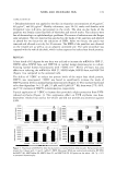
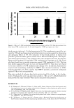
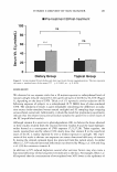
216 JOURNAL OF COSMETIC SCIENCE structure. In these models, cell migration via fibronectin connections, and cell multiplication are able to be observed and quantified. The results show that the selected active compound (Deliner®) induced a significant increase (p0.01) in the number of fibroblasts having colonized the artificial wound: + 21% as compared to the control. It also stimulated the cellular proliferation of fibroblasts in this area ( + 26% as compared to the control). Parallel to this stimulation of the re-colonization, used at 1%, this selected material increased fibroblastic fibronectin production in this area: + 56% as compared to the control (p0.05). A correlation could be foreseen between the different parameters measured. The increase in fibronectin quantities present in the Reconstructed Skin could induce cellular migration and proliferation, through the creation of a veritable intra-dermal "scaffolding" action. Selected active compounds therefore proposes, through the stimulation of cutaneous fibronectin production, an increased fibroblastic re-colonization of areas from which these cells had been eliminated. This re-colonization logically should result in the origin of an extracellular new matrix formation, COLETICA chose to evaluate the anti-wrinkle effects of DELINER® in an jn vjvo study on healthy volunteers. 20 healthy female volunteers have been used for this study performed on a "half-face". During 56 days, each volunteer applied on a "half-face" a placebo formulation and a formulation containing 3% of the selected active compound on the other "half-face". Before and after the 56-day treatment, the wrinkle depth of each "crow's feet" of volunteers was performed without modifying the volume of the analysed area using a "fringe projection" analysis method. The results show that, a 56-day treatment was able to significantly improve the cutaneous relief of the volunteers on the "crow's feet" area: depth of main wrinkles -7.9% (ns), micro-depressionary network (wrinkles and lines) -11.2% (p0.05), lines -13.5% (p0.05). Under the same experimental conditions, the placebo formulation did not induce any significant reduction of the measured parameters: depth of main wrinkles -3% (ns), micro-depressionary network (wrinkles and lines) -0.3% (ns), lines only +1.8% (ns). In conclusion, the selected active compound through perfectly identified metabolic effects, offers a significant reduction in wrinkle depth on the "crow's feet" area of volunteers. From this work first, it must be agreed today that certain messages from keratinocytes regulate fibroblastic fibronectin production throughout the life cycle. Moreover, some active compounds are capable of acting on all of the phenomena acting during the wound-healing process, by stimulating fibroblastic fibronectin production. In addition to this specific action on the pathological process targets, such active compounds could reorganizes the macromolecules of the extra-cellular matrix, the only controller of concerted cellular adhesion, movement and migration within skin tissue. In a particularly relevant way in term of cutaneous physiology, the specific metabolic activities of one of this selected ingredient result in vivo in a significant "anti-wrinkle" effect. Bibliography KARTTUNEN T., RISTELI J., AUTIO-HARMAINEN H., RISTELI L., Effect of age and diabetes on type IV collagen and laminin in human kidney cortex, Kjdney Int. 30, 586-591 (1987) OIKARINEN A., Aging of the skin connective tissue: how to measure the biochemical and mechanical properties of aging dermis, Photodermatol. Photojmmunol. Photomed. 10, 47-52 (1994) PEACOKE M., CAMPISI J., Cellular senescence: a relation of control, differentiation or aging?, J. Cell 8jochem. 45, 147-155 (1991) PIERAGGI M., JULIAN M., BOUISSOU H., Fibroblast changes in cutaneous ageing, Vjrchows Arch. A. Pathol. Anal. Hjstopathol. 402, 275-287 (1984) RUOSLAHTI E., Proteoglycans in cell regulation, J. 8jo/. Chem. 264, 13369-13372 (1989) TSUJI T., Ultrastructure of deep wrinkles in the ederly, Cutan. Pathol. 14, 158-164 (1987) UITTO J., Connective tissue biochemistry of the aging dermis. Age-related alterations in collagen and elastin, Dermatol. Clin. 4, 433-446 (1986)
Purchased for the exclusive use of nofirst nolast (unknown)
From: SCC Media Library & Resource Center (library.scconline.org)