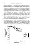
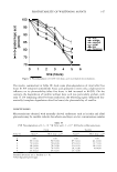
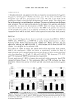
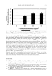
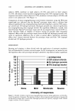
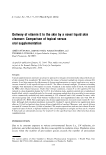
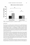
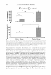
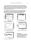
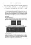
2003 ANNUAL SCIENTIFIC MEETING 223 Release of tested absorbed organic species (C20H23N·HC1) was obtained by limited dilution and the results are plotted in Figure 3. When equilibrium is reached, 20% of the drug is released, indicating the nanogels to be good carriers to encapsulate small molecules and to have the potential for controlled release applications. In Figures 4 and 5, kinetics of small molecule binding and release processes was investigated using surface plasma resonance spectroscopy (SPR), a powerful technique for monitoring short-term and long-term changes. Results obtained are shown in Figures 4 and 5. Both processes are quick and the equilibrium is established within 20 minutes. These nanogels, as a type of carriers, exhibit flexibility for tailoring for different applications. By combining the effects of the crosslinking density and functional groups, nano gels can provide efficient delivery and release of active molecules. The build-up of structure-property relationships between functional nanogels and organic molecules provides a powerful means for designing carriers for uptake/release of fragrances as well as other types of active components with nano gels. 350 1, 300 i 250 � g 2 00 .. §] 150 i! � 100 1 50 a Nanogel Concentration = 5 mg/ml, In water \. C 20 H 13 N · HCI Concentration = 0.5 mg/ml 6-----.C6/Neg -·- -A._____________, C6 Neg l,_11---Un_m_o�dlfl_ed_····--·-·-··-··-t 0 +----,----.----.----,----,----1 0 10 12 Crosslinking Density,% Figure 1. C2oH23N·HCl binding with nanogels as a function of crosslinking densities 80 100 .. ::,7 80 t 60 ·a .. 60 � 40 40 "2 B 2 0 'a .. ' " · - · - •···II•,. - - II 20 � 0 0 0 100 2 00 300 400 500 Bound Active by N anogels, micromole Figure 3. CzoH23N·HCl release from nanogels C 20 H13N·HCI, Temp.= 25'C, pH= 6 .0.02 _____________ __, 20 40 60 80 100 110 1 Time, minutes Figure 5. Kinetics of C2oH23N·HCl release � '$. ii 1 � � � 0,82 0.78 0.74 0.7 0.66 0,62 0.58 C6nanogels ./_,. .,. , .,... Urmodffled 0.5 1.5 Nanogel Coo.centratim, wt% 2,5 Figure 2. Pyrene encapsulation with Nanogels 0.00� C 2 off23N·HC�25-C, pH= 6 20 40 60 80 100 120 140 Time,minute Figure 4. Kinetics of C2oH23N·HCl binding Acknowledgements We acknowledge the supports of National Science Foundation Engineering Research Center for Particle Science and Technology at University of Florida and National Science Foundation Industry/University Research Center for Advanced Surfactants at Columbia University

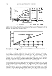
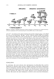
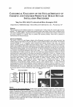
224 JOURNAL OF COSMETIC SCIENCE ADVANCED HISTOLOGICAL TECHNOLOGY AND THREE-DIMENSIONAL IMAGING: A FRUITFUL PARTNERSHIP TO VISUALIZE THE HUMAN SKIN MICRO-ANATOMY AND CHANGES IN CUTANEOUS STRUCTURES Gilles Pauly, M.D., Marie-Danielle Vazquez-Duchene, Ph.D., Dominique Gauche, Jean-Luc Contet-Adooneau, M.D., Christine Jeanmaire, Ph.D., Louis Danoux and Olga Freis, Ph.D. Laboratoires Serobiologiques, Division of Cognis, France, Pulnoy, France In our laboratories, from classical histology and immunohistochemistry to in situ hybridization with the use of photonic, mono and two photons confocal systems, morpho-functional studies allow to explore into a 3D space internal and external human cutaneous structures [ 1 ). Object velocity and acceleration in movement into a 3D space require eyes nimbleness for following displacements. Repetitive sequences in mo\ie facilitate understanding of biological mechanisms [2]. Therefore, 3D representation gives us a schematic inlerpretation of structural aspect more peninent than 2D images. To link 3D representation and cosmetology is an original approach for identifying structural cutaneous elements, for following up morphological changes due to intrinsic (age, gender, ... ) or extrinsic factors (UV, pollution, temperature, ... ) and for \'isualizing cosmetic effect. Which assets can bring 3D representation to histology and cosmetology? The detail is infinitely small. Let's jump to explore the intriguing world of skin. Look on epidermalcells Apoptosis localization inside keratinocytes nuclei Skin homeostasis is linked with apoptosis activity which is re\'ealed by TUNEL technique on keratinocytes nuclei (green in Fig. l). Skin sample was investigated by confocal microscopy. Then, 3D biological objects (red and green channels for skin and nuclei respecti\'ely) were obtained 10 show the nuclear density, their spreading on face (Fig. I a) and profile (Fig. I b). We can apply this technology after treatment on skin by an anti-aging active ingredient. Fig. 1: Apoptosis revealed by TUNEL technique on keratinocytes (a: at 0° and b: at 30° rotation). The use of two-photons microscope allows 10 penetrate deeper into the skin. Transparency effect, volumic rendering (Fig. 2a), targeted magnifying and rotation (Fig. 2b to 2d) provide a new glance on keratinocytes nuclei for analyzing apoptosis spreading. Fig. 2 : View on number, spreading and location of nuclear apoptosis by volumic rendering, magnifying and rotation. Extraction of dendritic epidermal cells Melanocytes and Langerhans cells, two types of dendritic cells present in the epidermis could be visualized by immunohistochemistry inside epidermis using \irnentin antibody. Vimentin (in green) recognizes intennediate filaments expressed by dendritic cells(Fig. 3). Computerized tool allows us 10 extract chosen cells with its epidermal context and to show another point of view. This way offers to examine cells from every angle. Cellular reconstruction imparts to the epidermis a living aspect. Pictorial artifices give a way for structures exploring. 3D visualization brings a superior dimension compared to the used traditional methods (INCi Name or LS active ingredient : Arginine (and) Mannitol (and) Disodium Adenosine Triphosphate (and) mannitol (and) Pyridoxine HCI (and) RNA (and) Histidine HCI (and) Phenylalanine (and) Tyrosine).
Purchased for the exclusive use of nofirst nolast (unknown)
From: SCC Media Library & Resource Center (library.scconline.org)