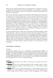
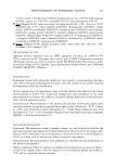
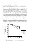
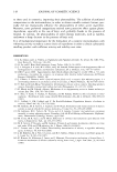
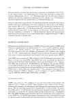
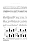
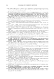
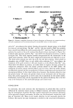
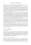
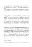
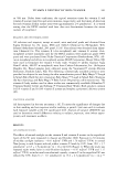
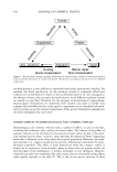
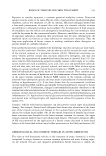
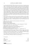
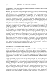
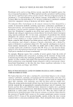
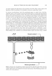
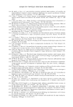
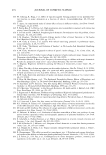
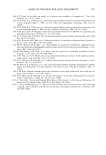
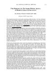
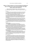
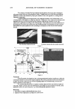
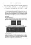
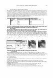
2003 ANNUAL SCIENTIFIC MEETING 225 Fig. 3: Extraction of Langerhans 's cell and melanocyte from epidennis ground. View on dermal components Glycosaminoglycans Fibroblasts are dennal cells which synthesize fibers and ground substance. By following the glycosaminoglycans spreading in fibroblasts, we can visualize distinctly a secretion area of chondroirin sulfate around the nucleus and the pericellular area (Fig. 4). Confocal microscopy acquisition gives us serial sections for reconstructing 3D fibroblastic representation and for measuring volume and area. Transparency effect on external fibroblastic "membrane" allows us to assess its secretion under LS Active Ingredient (INCi name: Mannitol (and) Cyclodwrin (and) Yea.�, Extract (and) Disodium Succinate). 3D rendering brings a functional benefit to micro-morphology developments. Fig. 4: Secretion area of chondroitin sulfate (in green) corresponds to cytoplasmic region around the nucleus (in red) and pericellular area which is made \.isible by the use of transparency effect. Elastic fibers The elastic fibers of the connective tissue form a network responsible for rhe elastic properties of skin. The organization of its network is not easily perceived with 2D images (3, 4]. The use of JD representation (Fig. 5), by a volumic rendering, allows to visualize the network organization, and the connection existing between elastic fibers. This volumic rendering corresponds to full elements, in opposition to surfacic rendering that shows only the external surface of those elements. JD visualization brings a superior dimension compared to the used traditional methods ( INCi Name of LS active ingredient: Pisum Sativum ( Pea) Extract). Fig. 5: Elastic fibers representation and its organization. In conclusion, volumic rendering offers a new approach of cutaneous structures. Extraction, segmentation, thresholding and measurements of these structures bridge the gap between histology and JD computeril.Cd representation. Then, 3D visualization allows to take into account the whole structure of skin which organizes the biological activities of cells and therefore a better evaluation of the activities from cosmetic active ingredient. By this way, 3D cosmetic universe delivers answers to many cutaneous challenges. (1] Vazquez-Duchfne MD, GiUon V, Contet-Audonneau JL, Freis 0, Perie G, Jeanmaire C, Gauch� D and Pauly G, Skin Research and Technology (abstract 53, ISBS-ISSI Hamburg). 9, 185. (2003). [ll Duchowski A, Medlin E, Coumia N, Murphy H, Gramopadhye A, �air S, Vorah J, and Melloy B, Behavior Research Methods, Instruments and Computers, 34, 573-591, (2002). [3) Jeanmaire C, DanOWI Land Pauly G, British Journal of Dennatology, 145, 10-18, (2001). (4) Contet-Audonneau JL, Jeanmaire C and Pauly G, British Journal of Dermatology, 140, 1038-1047, (1999).

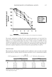
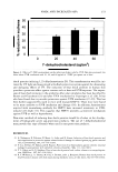
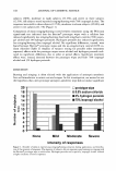
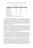
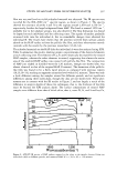
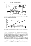
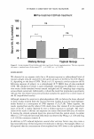
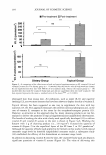
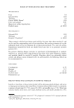
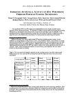
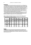
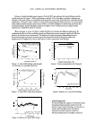
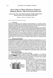
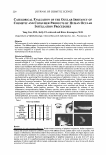
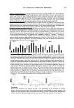
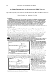
226 JOURNAL OF COSMETIC SCIENCE PUFFY EYES: A MULTI-FACTORAL COSMETIC PROBLEM NEEDS A MULTI-FACETED SOLUTION Karl Lintner, Ph.D., Claire Mas-Chamberlin, Philippe Mondon, Ph.D., Olivier Peschard, Ph.D. and Francois Lamy, Ph.D. Sederma, Le Perray en Yvelines, France Key words: lymphatic drainage, bradykinin, capillary fragility Introduction "Puffy eyes" are a well-known aesthetic problem for the ageing population. It is not possible to define a single cause for the slight swelling, the slackening of the skin, the occasional redness and sensitivity of the area immediately beneath the eyes where the skin is particularly thin (0.5mm on average). Several factors contribute to the emergence of bags under the eyes and their accentuation over time: a) CapillaIJ' fragility: The eyelid is crossed by a fine, dense network of arterial and venous capillaries. With age and outside influences, the vessel walls become fragile and plasma fluid leaks from the vascular bed b) Lymphatic drainage: The accumulation of interstitial fluids, when not eliminated by the lymphatic system, results in local overload and distension of the skin: defective drainage greatly contributes to the fonnation of bags under the eyes. c) Aging: The constant use of the eyelids and the fineness of their epithelium fairly rapidly result in tissue sagging with weakening of the supporting structure around the microvessels and in increases of dilated areas that retain edema. An inflammatory component generally accompanies infiltration of the tissues. The combination of those three factors is above all responsible for the phenomenon 'bags and rings under the eyes'. This paper describes an approach to treat these symptoms with a combination of several active substances intended to manage each of the major etiologies described above. Materials and Methods Hesperidine methyl chalcone is obtained by extraction and purification of hesperidine from citrus sinensis fruit, followed by selective methylation. The dipeptide Val-Trp (VW) and the tetrapeptide Pal-Gly-Gln-Pro Arg (Pal-GQPR) were obtained by liquid phase peptide synthesis and purified to 90%. Normal human keratinocytes were neo-cultured in standard medium until subconfluence, then exposed to increasing concentrations of Pal-GQPR IL6 secretion into the culture medium was quantified by ELISA method. In a second protocol, the cells were subjected to UVB,radiation (35 rnJ/cm2) after 24 hours of culture in the presence or absence of Pal-GQPR, then cultured with fresh medium for a further 24 hours. Angiotensin converting enzyme and its substrate kit were obtained from SIGMA. A.C.E. inhibition was measured at A= 340 nm. Clinical study (in vivo) was carried out on 20 female volunteers with chronic "bags" under the eyes. Measurement of skin profile used the fringe projection method (EoTech, France) coupled to image analysis (lnnov Metric Software INC, USA). Standard selection criteria applied, Student's t-test for paired series was used to analyze the data. Results and Discussion: In vitro studies: a) inhibition of bradykinin metabolism The contraction of lymphangions, necessary to ensure drainage, is dependent on the endogenous nonapeptide brndykinin (BK) which activates the 02-receptors of the lymphangion (vasa lymphatica), and increases the frequency of contractions [1,2,]. However, the lifespan of BK is relatively short it is rapidly fragmented by a degradation enzyme, angiotensin converting enzyme or A.C.E [4]: A.C.E, through its action, thus controls the frequency and intensity of the lymph pulsations. The dipeptide Val-Trp (VW) is a natural fragment of enzymatic hydrolysis of food proteins and was 100 BO 60 40 20 FAPGG Fig.1 Bnldyklnln described by Saito [3] as occurring in fermented sake and sake lee. It is a potent inhibitor of the angiotensin converting enzyme: incubation of the A.C.E. with a synthetic substrate (furyl-aryloyl-phenylalanyl-gly gly=F APGG) leads to release of F AP and GG fragments and a measurable UV signal. Figure I shows the concentration dependent inhibition of this reaction. Figure I shows also the inhibition of the bradykinin breakdown by Val-Trp. Inhibiting the metabolism of bradykinin, increasing the local BK pool is thus intended to stimulate the pumping frequency of the lymphatic canals and to improve drainage.
Purchased for the exclusive use of nofirst nolast (unknown)
From: SCC Media Library & Resource Center (library.scconline.org)