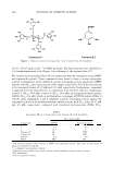
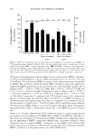
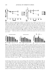
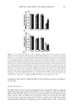
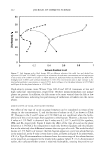
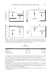
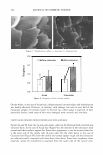
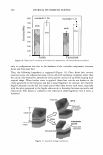
292 JOURNAL OF COSMETIC SCIENCE areas of the face and the neck (3,4). In addition, maintaining a uniform and lighter basal skin tone is a global concern that is a result of personal preferences and/or cultural biases. Due to the therapeutics and socio-economic importance of altering skin pigmentation, many efforts have been made to develop and recognize compounds that act as depig menting agents. Various remedies are available in the market, but none are completely satisfactory (4). Therefore, the development of an effective, controllable, and safe agent to regulate melanin synthesis in the skin is of great interest to medical practitioners and their patients. As a result of the key role played by tyrosinase in melanin biosynthesis, most popular skin depigmenting products use a tyrosinase inhibitor as an active ingredient (e.g., hydroquinone, kojic acid, arbutin). The success of treatment using these products is limited for two basic reasons: safety or overall effectiveness. Hydroquinone (HQ) has been banned in cosmetic use in Europe and is currently available only by prescription. It is considered a melanocyte cytotoxic agent as a consequence of its oxidation, through tyrosinase and/or spontaneously, to highly reactive species such as hydroxybenzoquinone and p-benzoquinone toxic products capable of disrupting fundamental cellular processes (4-7). Kojic acid (KA), an inhibitor of tyrosinase, has high sensitizing potential (8,9) and recently has been banned in Japan because of mutagenecity concerns (10). The effectiveness of KA has been demonstrated both in vitro (11,12) and in clinical trials (13,14). However, a relatively recent study demonstrated that KA treatment is not associated with reduction in pigmentation, using a cell-based assay (15 ). Arbutin (AR) has been traditionally used in Japan to treat pigmentary disorders (16,17), but its effectiveness is controversial. While several studies have demonstrated that arbutin can inhibit tyrosinase activity in cultured human melanocytes (17 ,18), many studies have demonstrated that arbutin increases pigmentation (19,20), or ineffectively inhibits ty rosinase activity both in the cell-free and cell-based assays (15 ). Because of the need to provide a skin depigmenting agent that is more efficacious, more stable, and less cytotoxic than the available skin-depigmenting agents, a novel com pound, deoxyarbutin (dA) 4-{(tetrahydro-2H-pyran-2-yl)oxy }phenol, was developed us ing quantitative structure-activity relationships (QSAR) of tyrosinase inhibitors (1). In this report, we analyze the safety, effectiveness, and reversibility of dA on human melanocytes in comparison to the aforementioned tyrosinase inhibitors, hydroquinone, kojic acid, and arbutin. In addition, we demonstrate the skin lightening effect of dA on human skin. MATERIALS AND METHODS CHEMICALS AND SOL VENTS Hydroquinone (HQ), kojic acid (KA), arbutin (AR), and 4-tertiary butylphenol (TBP) were purchased from Sigma Chemical (St. Louis, MO). Deoxyarbutin (dA) was initially synthesized by the authors and scaled up by Girindus America Inc. (Cincinnati, OH). For culture studies, dA, HQ, KA, and TBP were dissolved in sterile DMSO, while AR was dissolved in MCDB 15 3 media (Irvine Scientific, Santa Ana, CA). TBP was included in this comparative study because it is a tyrosinase inhibitor implicated in contact/ occupational vitiligo (21). Stock solutions of compounds were prepared and protected

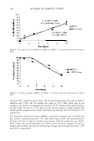
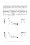
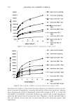
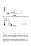
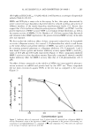
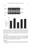
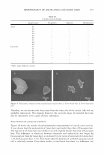
EFFICACY AND SAFETY OF DEOXY ARBUTIN 293 from light at -20°C until use. At the time of use, the compounds were further diluted in growth medium to the final concentration. For the animal study, 5 % dA, HQ and TBP were prepared in a mixture of propylene glycol, ethanol, and water at a volume ratio of 1:2:1. Solutions were kept at -20°C until use. CELL CULTURE Primary cultures of normal human melanocytes, keratinocytes, and fibroblasts were established from individual neonatal foreskins [from dark (dk) and light (lt) skin infants} that were obtained from the nursery of the University Hospital in Cincinnati after routine circumcision using a protocol approved by the University of Cincinnati Insti tutional Review Board as previously described (22). Foreskins were incubated in 0.25% trypsin for two hours at 37 ° C. The tissue was gently vortexed for 30 s to separate the dermis as a single piece and produce an epidermal cell suspension. The epidermal cells were seeded in a T-25 cm2 flask in either melanocyte or keratinocyte growth medium. Melanocytes were maintained in MCDB-15 3 growth media (Irvine Scientific, Santa Ana, CA) supplemented with 4% fetal bovine serum, 1 % antibiotic/anti-mycotic solution (Gibco, BRL, Grand Island, NY), 1 µg/ml vitamin E, 0.6 ng/ml human recombinant basic fibroblast growth factor, 5 µg/ml insulin, 0.05 µg/ml transferrin, 13 ng/ml bovine pituitary extract (Clonetics, Walkersville, MD) and 8 nM 12-O-tetradecanoylphorbol- 13-acetate. All of the above reagents were from Sigma Chemical Co. (St. Louis, MO) unless otherwise stated. The growth medium for normal human keratinocyte cultures consisted of Ml 54 basal medium (Cascades Biologicals, Portland, OR) supplemented with human keratinocyte growth supplement (Cascade Biologicals) and 1 % antibiotic/ anti-mycotic (Gibco). The dermis was vortexed and seeded into a T-25 cm2 flask with fibroblast growth medium consisting of DMEM medium (Gibco) containing 8% fetal bovine serum, 1 % glutamine (Gibco), 1 % sodium pyruvate (Gibco) and 1 % antibiotic/anti-mycotic solu tion (Gibco). All cultures were maintained in a tissue culture incubator at 3 7 ° C with 5 % CO 2 . The growth medium was routinely changed twice a week for melanocytes and fibroblasts and every other day for keratinocytes. CELL VIABILITY ASSAY Cell number (i.e., viability) was determined by direct counting. In this technique, cells were seeded at 1.3 x 10⁵ cells for melanocytes, 5 x 10⁴ cells for fibroblasts, and 5.5 x 10⁴ cells for keratinocytes, per T-12.5 cm2 flask. Cells were allowed to attach and grow for 48 hr before treatment. Cells were then treated daily with fresh growth media containing test compounds for five days. Compounds were tested at a range of concen trations in order to determine the maximum dose that did not affect the viability of human cells (experiments were performed at least in duplicate). On the sixth day, cells were detached with lX trypsin/EDTA and counted with a Coulter Counter. To deter mine the fraction of cells surviving the treatment in a specified concentration of an experimental agent, the viable cell number after each treatment was normalized using the average of the viable cell number in a control group.
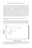
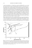
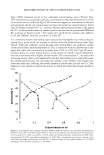
Purchased for the exclusive use of nofirst nolast (unknown)
From: SCC Media Library & Resource Center (library.scconline.org)