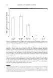
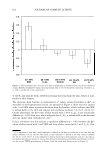
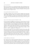
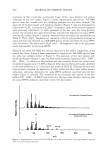
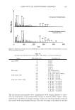
]. Cosmet. Sci.) 58, 599-620 (November/December 2007) Ranking of aqueous surfactant-humectant systems based on an analysis of in vitro and in vivo skin barrier perturbation measurements SASWATA GHOSH, SIDNEY HORNBY, GARY GROVE, CHARLES ZER WICK, YOHINI APP A, and DANIEL BLANKSCHTEIN, Department of Chemical Engineering, Massachusetts Institute of Technology, Cambridge, MA 02139 (S.G., D.B.)) Neutrogena Corporation, 5760 West 96th Street, Los Angeles, CA 90045 (S.H., Y.A.), and Cyber DERM Clinical Studies, 700 Parkway Drive, Broomall, PA 19008 (G.G., C.Z.). Accepted for publication July 19, 2007. Synopsis We propose that skin electrical current measurements can be used in vitro to effectively rank aqueous solutions containing surfactants and humectants (the enhancer) contacting the skin, relative to a PBS aqueous solution (the control) contacting the skin, based on their ability to perturb the skin aqueous pores. Specifically, we develop an in vitro ranking metric using the increase in the skin electrical current induced by an enhancer relative to the control. Aqueous contacting solutions containing (i) surfactants [SDS (sodium dodecyl sulfate)} and C 12 E6 [dodecyl hexa (ethylene oxide)}, (ii) humectants (glycerol and propylene glycol), and (iii) a control (PBS) were studied. Utilizing the new in vitro ranking metric, these aqueous contacting solutions were ranked as follows (from the mildest to the harshest): glycerol propylene glycol PBS C 12 E6 SDS. In order to further develop this ranking methodology, which can potentially lead to the reduction, or elimination, of costly and time-consuming procedures, such as human and animal testing and trial-and-error screening in vivo, it was important to correlate the findings of the in vitro ranking metric with direct in vivo skin barrier measurements. For this purpose, in vivo soap chamber measurements, including transepidermal water loss, visual skin dryness, and chromameter erythema measurements, were carried out on human volunteers using the aqueous surfactant-humectant solutions described above. The results of these in vivo measurements were found to be consistent with the ranking results obtained using the in vitro ranking metric. To further explore the validity of our model and to verify the skin barrier mitigating effect of glycerol, in vivo soap chamber measurements were carried out for aqueous SDS solutions containing 10 wt% added glycerol. These in vivo measurements support our recent in vitro finding that glycerol reduces the average radius and the pore number density of the skin aqueous pores, such that SDS micelles are hindered from penetrating into the skin and inducing skin barrier perturbation. INTRODUCTION The stratum corneum (SC) serves as the barrier between the body and the environ- Address all correspondence to Daniel Blankschtein. 599

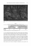
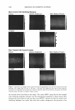
600 JOURNAL OF COSMETIC SCIENCE ment (1). Compared to the porous structure of the viable epidermis and the porous and hydrated structure of the dermis, which lie beneath the SC, the rigid and ordered structure of the SC is primarily responsible for the skin barrier function (2,3). The SC possesses an ordered brick-and-mortar structure, which consists of the flat corneocytes (the cellular bricks), interlocked with the lipid lamellae (the intercellular mortar) (1-3). The lipid lamellae of the SC consist of lipid bilayers alternating with aqueous, hydro philic layers (1-4). The observation that hydrophilic solutes are able to permeate across the SC, even under passive skin permeation conditions, has led researchers to propose the existence of tortuous, aqueous pores through the intercellular lipid lamellae in the SC. In fact, Menon and Elias (4) have established a morphological basis for the existence of aqueous pores in the mammalian SC. By applying hydrophilic and hydrophobic tracers in vivo to murine skin, under passive skin permeation conditions and under enhanced skin permeation conditions including chemical enhancers, sonophoresis, and iontopho resis, Menon and Elias visualized the resulting penetration pathways using ruthenium tetroxide staining and microwave post fixation methods (4). These aqueous pores in the stratum corneum (SC) are located in the lacunae and other aqueous regions, surrounded by polar lipids in the lipoidal mortar between the corneocyte bricks that comprise the brick-and-mortar structure of the SC (4-9). In fact, diffusion through aqueous pores in the SC has been used to explain skin penetration and the subsequent permeation of hydrophilic permeants, including mannitol (5-9), current-carrying ions like Na + and Cl - (5 ,9), and micelles of surfactants like sodium dodecyl sulfate (SDS) ( 6,29). Macroscopic in vitro skin barrier measurements, including average skin electrical resis tivity (R), which is a measure of the permeability of the current-carrying ions, and mannitol transdermal permeability (P), have been used in the context of a hindered transport theory to calculate the average pore radius (rpore) and the porosity-to-tortuosity ratio (e!T) 1 of the skin aqueous pores of the SC (5-9). Specifically, it is possible to quantify the extent of in vitro skin barrier perturbation that an aqueous solution con sisting of surfactants2 and humectants3 induces by examining the modifications of 'pore and e/T relative to an in vitro control such as PBS (phosphate-buffered saline) (5-9). The harsh surfactant SDS was shown to induce significant skin barrier perturbation in vitro relative to the PBS control, while the humectant glycerol was shown to preserve the skin barrier in vitro and to mitigate skin barrier perturbation ( 6, 12-16,21-24,30-3 5 ). Classical bioengineering techniques have been used extensively (12-16,21-24,30-35) to assess and rank the impact of surfactant solutions on human skin. However, these methods provide limited understanding of the mechanism of skin barrier perturbation. 1 The porosity, s, of the aqueous pores is defined as the fraction of cross-sectional SC area occupied by the aqueous pores, and the tortuosity, 'T, is defined as the ratio of the tortuous length of the aqueous pore to the thickness of the SC (5-9,10,11). 2 Surfactants are surface-active agents that are commonly used in skin cleansing formulations because of their ability to stabilize oil-water emulsions and clean the surface of the skin. However, they may penetrate into the skin and induce skin barrier perturbation (6,12,13,16,21,22,29). They have a hydrophilic head and a hydrophobic tail, and self-assemble to form micelles at a concentration above the critical micelle concen tration (CMC). 3 Humectants maintain the natural water content of the skin and preserve the skin barrier (30-3 5 ). In addition, some humectants like glycerol have been shown to mitigate surfactant-induced skin barrier perturbation in vitro, by preventing micelles of surfactants like SDS from penetrating into the skin through aqueous pores (6).
Purchased for the exclusive use of nofirst nolast (unknown)
From: SCC Media Library & Resource Center (library.scconline.org)