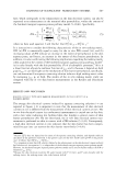
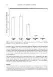
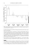
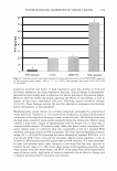
664 JOURNAL OF COSMETIC SCIENCE of nails. A liquid nail treatment formulated with panthenol efficacy and good tolerance of C8-LHA make it an (2%) was compared to a solution of panthenol (2%) in water. excellent candidate for the treatment of hyperpigmentory Fingernail specimens were dosed daily for 7 days with either disorders. the nail treatment (non-lacquer film forming) formulation or aqueous solution with sampling performed every 24 h. Panthenol concentrations were determined in the dorsal surface, interior (by drilling and removal) and in the supporting bed under the human nail. Panthenol levels in the dorsal nail (R2 = 0.87 P 0.001), nail interior (R2 = 0.94 P 0.001) and nail supporting bed (R2 = 0.79 P 0.003) showed a significant linear increase with each day of dosing. Significantly more panthenol was delivered into the interior nail and supporting bed by a nail treatment formulation than from an aqueous solution. The film acts not only as a reservoir of panthenol, but also acts to increase the hydration of the nail and the thermodynamic activity of panthenol as well, thereby enhancing diffusion A Simple Experimental Method To Study Depigmenting Agents M. L. Abella, J. de Rigal and S. Neveux L'Oreal Recherche, Chevilly-Larue, France Dr M. L. Abella, Centre de Recherche L'Oreal, 188 rue Paul Hoebart, 94550 Chevilly Larue, France. The first objective of the study was to verify that a controlled UV exposure of four areas of the forearms together with randomized product application enabled to compare treatment efficacy and then to compare the depigmenting efficacy of different products with a simple experimental method. Sixteen volunteers received 0.7 minimal erythermal dose for four consecutive days. Products tested were ellagic acid (0.5%), vitamin C (5%) and C8-LHA (2%). Product application started 72 h post last exposure, was repeated for 42 days, the control zone being exposed, non-treated. Colour measurements included Chromameter®, Chromasphere®, Spectro-colorimeter and visual assessment. Comparison of colour values at day I and at day 7 showed that all zones were comparably tanned, allowing a rigorous comparison of the treatments. We report a new simple experimental model, which enables the rapid comparison of different depigmenting products. The Review Article: Nail Biology And Nail Science D. A. R. de Berker*, J. Andret and R. Baran* *Bristol Dermatology Ce r, e, Bristol Royal Infirmary, Bristol BS2 8HW, U.K., Department of Dermat � ogy, CHU Saint Pierre, 1000 Brussels, Belgium and Nail Disease Centre, 42, rue des Serbes, 06400 Cannes, France Robert Baran, Nail Disease Centre, 42, rue des Serbes, 06400 Cannes, France The nail plate is the permanent product of the nail matrix. Its normal appearance and growth depend on the integrity of several components: the surrounding tissues or perionychium and the bony phalanx that are contributing to the nail apparatus or nail unit. The nail is inserted proximally in an invagination practically parallel to the upper surface of the skin and laterally in the lateral nail grooves. This pocket-like invagination has a roof, the proximal nail fold and a floor, the matrix from which the nail is derived. The germinal matrix forms the bulk of the nail plate. The proximal element forms the superficial third of the nail whereas the distal element provides its inferior two-thirds. The ventral surface of the proximal nail fold adheres closely to the nail for a short distance and forms a gradually desquamating tissue, the cuticle, made of the stratum comeum of both the dorsal and the ventral side of the proximal nail fold. The cuticle seals and therefore protects the ungual cul-de-sac. The nail plate is bordered by the proximal nail fold which is continuous with the similarly structured lateral nail fold on each side. The nail bed extends from the lunula to the hyponychium. It presents with parallel longitudinal rete ridges. This area, by contrast to the matrix has a firm attachment to the nail plate and nail avulsion produces a denudation of the nail bed. Colourless, but translucent, the highly vascular connective tissue containing glomus organs transmits a pink colour through the nail. Among its multiple functions, the nail provides counterpressure to the pulp that is essential to the tactile sensation involving the fingers and to the prevention of the hypertrophy of the distal wall tissue, produced after nail loss of the great toe nail.

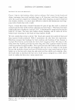
ABSTRACTS 665 Salicyloyl-Phytosphingosine: A Novel Agent For The Repair Of Photoaged Skin M. Farwick*, R. E. B. Watson f, A. V. Raw y ngs *, U. Wollenw t r*, P. Lersch*, J 't J. Bowden , J. Y. Bastrilles and C. E. M. Griffiths *Degussa AG, Goldschmidt Personal Car f Goldschmidtstrasse 100, 45127 Essen, Germany, Dermatopharmacology Unit, Dermatology Centre, Ho 'f Hospital, The University of Manchester, Manchester and A VR Consulting Ltd, Northwich, Cheshire, U.K. Mike Farwick, Degussa AG, Goldschmidt Personal Care, Goldschmidtstrasse 100, 45127 Essen, Germany In recent years the importance of sphingolipids ( cerebrosides, sphingomyelin, ceramides, sphingosine-1- phospate, etc.) in skin biology is receiving an increasing interest. Not only are ceramides essential for the barrier function of the skin, especially through their phytosphingosine, sphingosine and 6-hydroxysphingosine derivatives, they are now also known to be cell-signalling mediators which can improve epidermal differentiation. However, their effects on dermal anti-ageing markers and reduction of wrinkles have not been established. In this study, we were interested in the effects of a sphingolipid derivative, salicyloyl-phytosphingosine (SP), because of the known independent beneficial effects of salicylic acid and phytosphingosine on skin. Both of these agents are known to reduce the activities of the activator protein-I transcription factor, in a manner similar to that observed with retinoic acid (RA) treatment. Through this mechanism, RA was shown to reduce the levels of matrix metalloproteases (MMPs) and the increase levels of extracellular matrix proteins. Therefore, we examined the effects of SP on procollagen-1 synthesis in fibroblasts in vitro, its effects in vivo on the expression of dermal markers such as fibrillin-1, procollagen-1 and MMP-1 immunochemically in biopsies taken from a short-term occluded patch test protocol and, its effects on periorbital wrinkle reduction over 4 weeks using Fast Optical In Vivo Topometry of Human Skin. In vitro we observed a significant increase in the production of procollagen-I by adult human fibroblasts (two fold increase, P 0.01) which encouraged us to test the effects of SP in vivo. Initially, test products (SP at 0.05% and 0.2%, all-trans RA (0.025%) and vehicle were applied under occlusion for 8 days prior to biopsy and histological assessment in photoaged volunteers (n = 5). Increased deposition of fibrillin-1 and procollagen-I, together with reductions in the levels of MMP-1, were observed for the SP treatments (P 0.05). Similar effects were observed for RA, except for the increases in procollagen-1. With these beneficial effects on the basement membrane and papillary dermal markers, we evaluated the effects of SP in an oil-in-water (0/W) cream for its effects in reducing the appearance of periorbital wrinkles in a 4-week, half-face clinical study compared to placebo cream (moderately photoaged female subjects aged 41----69 years n = 30). Clear reductions in wrinkle depth and Rz (skin smoothness) together with Ra (skin roughness) parameters were observed (P 0.05), indicating an anti wrinkle benefit. In conclusion, this series of studies demonstrated for the first time that a ceramide derivative, such as that SP, was a novel agent for the repair of photoaged skin and highlight its effects at the cellular, tissue and organ levels. Three-Dimensional Imaging And Analysis Of The Surface Of Hair Fibres Using Scanning Electron Microscopy C. Tomes*, J. T. Jones*, C. M. Carr" and D. Jones f *Textiles and Paper, School of Materials, Th f University of Manchester, Manchester M60 1 QD and Croda Chemicals, Cowick Hall, Goole DN14 9AA, U.K. Chris M. Carr Tomes, Textiles and Paper, School of Materials, The University of Manchester, Manchester M60 1QD,U.K. Cuticle scales are the most obvious feature of a hair fibre's outer surface and much research has focused on assessing the influence of surface topography on the associated hair fibre's properties. However, much of the research has either been qualitative or, if quantitative, employed relatively laborious analytical techniques to establish the necessary statistical robustness. In this study, we report on the application of a 3D image analysis package capable of producing 3D data from multiple 2D scanning electron microscope (SEM) images of hair fibres. Analysis of the surface profile can be carried out quickly and accurately, enabling quantification of the scale structure. To validate the novel technique and ensure that the scale heights measured were indeed accurate and reproducible, extensive calibration of the SEM and the 3D software has been performed. In addition, scale heights on a single hair have
Purchased for the exclusive use of nofirst nolast (unknown)
From: SCC Media Library & Resource Center (library.scconline.org)