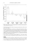
RANKING OF SURFACTANT-HUMECTANT SYSTEMS 601 Indeed, it would be of value to be able to correlate observations of the extent of skin barrier perturbation observed in vivo and the modification of the skin aqueous pores that an aqueous surfactant-humectant system induces in vitro relative to a control. This would allow one not only to rank these systems based on their ability to perturb the skin aqueous pores in vitro, but also to understand the mechanisms of perturbation. With the above in mind, we have developed an in vitro ranking metric that combines the characteristics of the skin aqueous pores and can potentially reduce, or eliminate alto gether, costly and time-consuming in vivo procedures such as testing for irritation potential and trial-and-error screening. Moreover, such a ranking metric would allow one to simultaneously screen and rank many surfactants and humectants for use in skin care formulations, thereby significantly speeding up the effort and time required to bring new skin care formulations to the market. In addition, a suitable ranking metric developed by measuring skin electrical currents induced in vitro relative to an in vitro PBS control can be combined with corresponding mannitol skin permeability measure ments, in the context of a hindered-transport aqueous porous pathway model. Specifi cally, use of the hindered-transport model enables the quantification of the modifications of: (i) the average pore radius, and (ii) the pore number density, induced by aqueous surfactant-humectant systems relative to the in vitro PBS control, thereby shedding light on the mechanism of in vitro skin barrier perturbation induced by the aqueous surfac tant-humectant system evaluated (see the Theoretical section). For this purpose, the anionic surfactant SDS and the nonionic surfactant C12E6 [dodecyl hexa (ethylene ox ide)}, and the humectants propylene glycol (PG) and glycerol (G), were selected (see the Materials section). The differences in the average pore radii and pore number densities induced by these two surfactants and two humectants relative to the in vitro PBS control were analyzed to gain mechanistic insight into their skin barrier perturbation/mitigation characteristics. Knowledge of the mechanism of in vitro skin barrier perturbation in duced by aqueous surfactant-humectant systems can be practically valuable in designing skin care formulations containing these chemicals that are mild to the skin and that minimize, or eliminate altogether, skin barrier perturbation (22-24,28,29,33,3 7). SDS is known to induce skin erythema (13-16,22), while C 12 E6 is known to induce skin dryness when applied to human skin in vivo (21,23,24). On the other hand, PG and G are both humectants, which are known to preserve the skin barrier and to maintain the water content of the skin when applied to human skin in vivo (30-36). The ranking of the two surfactants and the two humectants relative to the in vitro PBS control was compared with various in vivo skin barrier measurements, and the correlation between the in vitro ranking metric analysis and the in vivo skin barrier measurements was investigated. To assess the condition of the skin barrier in vivo, a soap chamber test, using a well accepted and previously published protocol (17 ,18,25-28), was utilized to treat the skin of the volar forearms of healthy human volunteers with the various surfactant-humectant solutions. Subsequently, the condition of the skin barrier was assessed by (a) transepi dermal water loss (TEWL) measurements to determine the moisture vapor flux over the skin as measured by an evaporimeter, which serves as an excellent quantitative indicator of skin barrier perturbation in vivo (25-27), (b) visual skin dryness scores determined by an expert grader to clinically assess the extent of skin dryness (17 ,23 ), and (c) skin erythema (redness), objectively determined by chromameter measurements (18). The

602 JOURNAL OF COSMETIC SCIENCE results of these in vivo measurements are reported here, and have been compared with the in vitro measurements. To further validate the in vitro ranking metric, we compared the in vitro and in vivo skin barrier mitigation effects of adding glycerol to SDS aqueous contacting solutions (6). This comparison demonstrates the ability of glycerol to mitigate skin barrier perturba tion upon exposure to SDS solutions, which provides evidence of the ability of glycerol to prevent SDS micelles from penetrating into the SC and inducing skin barrier per turbation in vivo. This important finding is fully consistent with our recent finding on the ability of glycerol to hinder SDS micelle penetration into the skin in vitro by reducing the average radius and the number density of the skin aqueous pores (6). EXPERIMENT AL IN VITRO SKIN BARRIER STUDIES Materials. Sodium dodecyl sulfate (SDS) was purchased from Sigma Chemicals (St. Louis, MO). Analytical-grade glycerol (G) and propylene glycol (PG) were purchased from VWR Chemicals (Cambridge, MA). 3 H-radiolabeled mannitol was purchased from American Radiolabeled Chemicals (St. Louis, MO). Dodecyl hexa (ethylene oxide), C 12 E6, was purchased from Nikko Chemicals (Tokyo, Japan). All these chemicals were used as received. Water was filtered using a Millipore Academic water filter (Bedford, MA). Phosphate-buffered saline (PBS) was prepared using PBS tablets from Sigma Chemicals (St. Louis, MO) and Millipore filtered water, such that a phosphate concen tration of 0.01 M along with a NaCl concentration of 0.137 M was obtained at a pH of 7.2. Preparation of the solutions. For the in vitro skin barrier studies, the following aqueous solutions were prepared: (i) an anionic surfactant solution-SDS (1 wt%) (ii) a nonionic surfactant solution-C 12 E6 (1 wt%) (iii) a control solution-phosphate-buffered saline (PBS) (iv) a humectant solution-propylene glycol (PG) (10 wt%) and (v) a humectant solution-glycerol (G) (10 wt%). Preparation of the skin samples. Female Yorkshire pigs (40-45 kg) were purchased from local farms, and the skin (back) was harvested within one hour after sacrificing the animal. The subcutaneous fat was trimmed off using a razor blade, and the full-thickness pig skin was cut into small pieces (2 cm x 2 cm) and stored in a -80°C freezer for up to two months. The in vitro experiments involve contacting pig full-thickness skin (p-FTS) with aqueous surfactant-humectant and PBS control solutions i-v, and were performed according to previously published protocol (5 ,6,29). Mannitol trans dermal permeability measurements. Vertical Franz diffusion cells (Permegear Inc., Riegelsville, PA) were used for the mannitol skin permeability measurements (5,6,29). All the experiments were performed at room temperature (25°C). The p-FTS samples were mounted in the diffusion cells with the SC facing the donor compartments. Both the donor and the receiver compartments were filled with PBS, and the p-FTS samples were left to hydrate for one hour before the beginning of the experiment to allow the skin's initial barrier property to reach steady state (5,6,29). At this point, the skin electrical current across the p-FTS sample was measured, and only p-FTS samples with an initial skin current 3 pA were utilized in the mannitol skin permeability studies
Purchased for the exclusive use of nofirst nolast (unknown)
From: SCC Media Library & Resource Center (library.scconline.org)