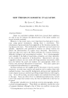
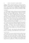
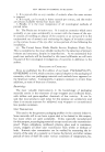
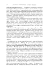
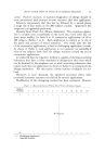
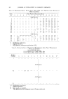
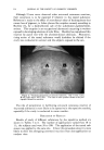
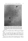
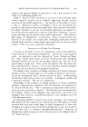
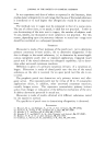
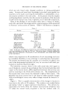
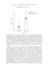
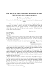
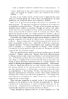
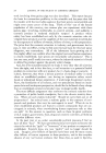
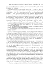
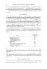
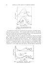
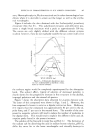
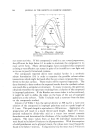
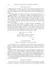
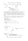
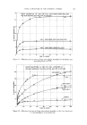
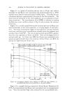
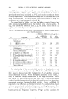
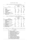
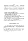
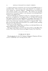
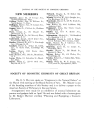
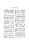
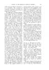
268 JOURNAL OF' THE SOCIETY OF COSMETIC CHEMISTS HYALURONIDASE ACTIVITY RANGE NORMAL PERIODONTAL DISEASE Figure 3. tissues. Two groups were selected (neither group contained any indi- viduals with upper respiratory disturbances). One group was composed of those who had no periodontal involvement and little or no caries experience. The other group contained individuals who had moderate to advanced periodontal involvement with little or no caries experience. Figure 3 illustrates the difference in hyaluronidase titers. The periodontally in- volved subjects demonstrated mean titers five to seven times that found in the mouth's of periodontally free individuals. These findings were first described by us in 1950 and 1952 (5, 11). Johnson, Chauncey and Lisanti reported findings in 1956 (12) which indicated that 80 per cent of the perio- dontally diseased and involved individuals showed abnormally high salivary protease activity, and that these titers remained high after therapy and 7O 6O "n 50 1• $O 20 H-'G- H'•: G- ENZYIVIES PRODUCED Figure 4. H-{': G+ H- NO HYALURONIDASE ACTIVITY G- NO $-GI.UCURONEIASE /$CTNIT• H+ HY/t&LJ•3NE)A•E ACTIVITY • $--GLUCURONII16SE ACTIVITY

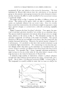
THE BEAUTY OF THE ENZYME SCREEN 269 healing had taken place. This is an indication of the potential of the bac- terial protease enzymes being involved in the cases of advanced periodontal disease. It has been well established as of the present date that the "so called" normal bacterial inhabitants of the oral cavity, and specifically Streptococ- cus mitis of the alpha hemolytic group as well as some of the staphylococcus and beta hemolytic streptococci are found in the oral cavity. These in- vestigations published in 1952 (5) and 1954 (13) indicate that many of these organisms produced both hyaluronidase and beta-glucuronidase and appear to be closely associated in the depolymerization of the inter-cement sub- stance found around and between cells. Figure 4 illustrates the enzyme characteristics of one type of oral isolate, Streptococcus mitis. Over 60 per cent of the Streptococcus mitis produced both hyaluronidase and beta glucuronidase, none were found which pro- duced beta glucuronidase alone, while 33 per cent produced hyaluronidase alone, therefore the postulation, that depolymerization of the mucopoly- saccharides of intact tissue may well be assisted by the action of hy- aluronidase and glucuronidase in a sequential manner. These findings from organisms normally found in the oral cavity may assist in the explanation of some of the nonspecific disease processes which have been previously as- sociated with the presence of certain "nonpathogens." We have been discussing en2 ymes from oral bacterial sources and one enzyme --•:• is ........ by the salivary glands and their relationship to •VIIILII oral and systemic disease conditions. In 1953 (14) we reported the presence of salivary cholinesterase which was found in both whole saliva and parotid saliva secretions. Several preliminary observations indicated that the parodd cholinesterase varied considerably and that this variation might be due to the attitude of the individual. Methods were developed to assay this enzyme activity in small quantities of parotid secretion and a study was undertaken to determine whether the enzyme titer was altered with the anxiety state or "stressed" state of the individual. Base lines were es- tablished for ten subjects over a period of three months for blood pressure, pulse rate and parotid cholinesterase titers. This was followed by a series of stress periods on these same subjects which consisted of taking blood, hyperventilation and cold pressors. The results demonstrated that both the anticipation of the "stresses" as well as the "stress" itself produced a depression of the mean cholinesterase levels while the blood pressure mean values did not change. Differences between the base line and the "stress" levels were statistically significant. These findings are illustrated in Fig. 5. On the left is the mean parodd cholinesterase before "stressed" state. The hash mark bar shows the drop in mean cholinesterase titer during the anx- iety or "stressful" state. As this graph indicates there was no change in pulse, diastolic or systolic pressure. These findings may in the future prove
Purchased for the exclusive use of nofirst nolast (unknown)
From: SCC Media Library & Resource Center (library.scconline.org)