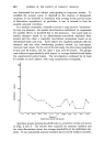
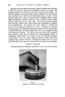
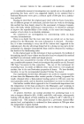
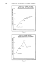
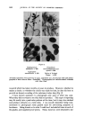
622 JOURNAL OF THE SOCIETY OF COSMETIC CHEMISTS These results showed coincident decrease in both P.ovale and dandruff in some individual cases but not in all in some cases, a fall in the dandruff index coincided with an increase in P.ovale in the early treatment period. Unfortunately, we did not manage to reduce infection virtually to nil for a prolonged interval and observe the clinical result. The findings from November 1962 to April 1963 may be regarded with suspicion owing to the abnormally cold weather. This was readily apparent in the P. ovale counts and was probably shown to some extent in the dandruff indices, in contrast to the untreated long-term series. It is also desirable to repeat the earlier suggestion that P.ovale may have an allergic rather than a toxic role if this were so, exacerbation and resolution would not necessarily be expected to coincide with infection and disinfection on a relatively short time-scale. HISTOLOGICAL STUDIES The lack of clear-cut evidence pointing to a micro-biological causation of dandruff suggested the need for histological study, which might have offered other possible mechanisms. For this purpose, it was essential to appreciate the dynamic nature of cellular activity in the epidermis and to regard the various layers seen in cross-section as representative of the changes taking place in depth. Epidermal cell-division is restricted mainly to the basal layer adjoining the dermis. In the stratum spinosum the precursors of keratin are first formed as tonofibrils and as granules of keratohyalin. The granules become more numerous and distinct as the cells move towards the surface, forming the stratum granulosum. Above this layer it is thought that the keratohyalin becomes converted to a fibrillar form in association with the tonofibrils. Orientation of the fibrils parallel to the surface may account for the bire- fringence of the next layer, the stratum lucidurn. Finally, the completely keratinized cells constitute the stratum corneum the horny cells retain little recognizable structure and have normally lost all traces of nuclear chromatin. The typical layers are easily demonstrable in the thick skin of the pall or sole but are not so clearly differentiated in the thinner epidermis of the scalp. In particular, the stratum granulosum is not continuous and may be difficult to detect in some tissue specimens. Stratum corneum normally desquamates as small, powdery particles. A Sellotape stripping of the outer skin shows that nothing but the outline of the cell shape remains. When dandruff is present, on the other hand, microscopic examination of the scales shows the presence of large amounts of nuclear chromatin. A cross-section of the scale shows that stratum corneum has been shed together with a large number of imperfectly keratin- ized cells still retaining their nuclei. This type of condition, in which the

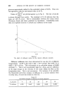
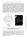
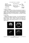
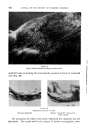
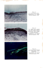
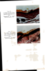
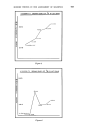
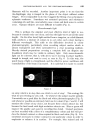
THE INVESTIGATION OF DANDRUFF 623 whole epidermis is thickened, and where nuclei persist in the stratum corneum, is known as parakeratosis. It is a usual feature of chronic in- flammation and seems to indicate conclusively that dandruff represents more than just an increase in the normal rate of exfoliation. An inflam- matory process does not necessarily imply itching or erythema, but is a response to some kind of irritant which may equally well be derived from a microbiological or immunological source, a chemical initiator or from psychogenic origins. It has proved just as difficult to identify the causative mechanism in the disease of psoriasis, where parakeratotic scaling bears sonhe resemblance to that seen in dandruff. Tickner (7) concluded that a metabolic block occurs in psoriasis, prevent- ing aggregation of the tonofibrils to form keratin. In our comparative studies, psoriatic scales were seen to contain cell-nuclei in all layers up to the surface, whereas dandruff scales usually retained some true stratum corneum. The psoriatic scales sometimes contained material staining like keratohyalin granules, but these were not seen in dandruff scales. Our observations do not suggest that a close analogy can justifiably be drawn between psoriatic scaling and dandruff, but further histological studies need to be undertaken for example, it would be interesting to seek evidence of micro-abscess formation {which is a diagnostic feature of psoriasis) by examination of biopsy material taken during acute episodes of dandruff when itching is pronounced. In the same circumstances, it would be helpful to know how deep within the epidermis the cells of P.ovale or other micro- organisms could be found. ANIMAL STUDIES No evidence of scaling in small animals has been described which would deafly indicate suitability for use in laboratory studies. However, it seemed reasonable to consider that, if dandruff was a parakeratotic response to low- grade irritation, an analogous situation should be capable of reproduction in an experimental species. This might, for example, be brought about by the topical application of oleic acid, which has been reported to cause epidermal hypertrophy and desquamation (8). Oleic acid applied to mouse skin produced gross irritation, but masking the carboxyl group by esteri- fication provided us with a suitable mild irritant. When ethyl oleate was applied to a region of growing hair on the mouse (C 57 BL strain), a response in the form of epidermal hypertrophy and parakeratotic scaling was obtained. The fur became filled with scales and the appearance resembled dandruff (Fig. 9). This persisted whilst appli- cations were continued and while the hair remained in the growing phase. During the resting phase of the hair growth cycle, epidermal response to ethyl oleate ceased. Ethyl oleate-induced scales bore a close similarity to
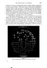
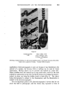
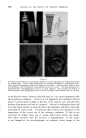
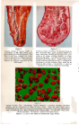
Purchased for the exclusive use of nofirst nolast (unknown)
From: SCC Media Library & Resource Center (library.scconline.org)