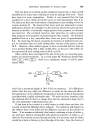
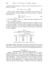
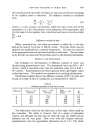
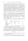
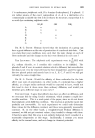
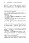
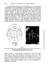
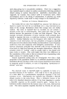
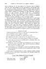
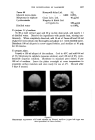
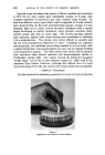
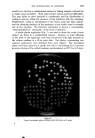
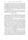
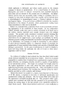
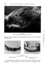
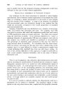
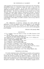
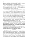
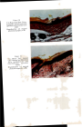
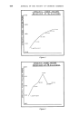
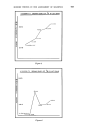
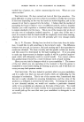
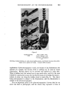
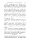
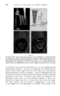
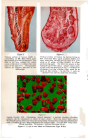
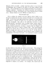
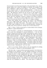
Figure 7 Vertical section of human eyelid in- corporating an 82C filter which gives the pleasant pale blue background. This filter in no way chan•es the colour values of the sta{n but colours the clear areas in the specimen. Photographed without an eyepiece attached to the microscope. Figure 8 Vertical section of human lip incorporating an 82A correction filter, again to give a better and more attractive background to that of a pure white or off white, so often revealed in colour photomicrography. The filter was placed in the light path, above the source. A large subject such as this, and that depicted in Fig. 7, is difficult to illuminate, and the extreme edges must be as sharp as the centres. As for Fig. 7, only an objective was used to make this recording. Figure .9 Aspirin Crystals X40. Illustrating "optical staining", a method whereby colourless subjects can be reproduced in colour of one's choice. Here the direct light was a Wratten 58 (green) filter providing the background, whereas the crystals were illuminated with a Wratten 29 (red) filter attached to the indirect light. Two 6V 30W lamps used. 25mm objective X4 eyepiece bellows extension 40 cm. Figures 7, 8, and .9 were made on Ektachrome Type B film.

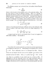
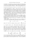
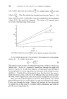
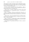
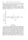
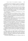
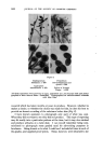
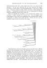
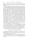
PHOTOMICROGRAPHY AND THE PHOTOMICROGRAPHER 649 filaments will be recorded. Another important point is to see that the iris-diaphragm stop is imaged in the plane of the object without colour fringes. If it is impossible to do this I suggest the fitting of an achromatic/ aplanatic condenser. Sometimes wet mounted specimens and substances containing oils and fats, unavoidably cause colour and dark zones in certain areas. Opaque subjects are more difficult to handle (Fig. $). Monochromatic light This is perhaps the simplest and most effective kind of light to use, because it contains only one colour, and thus the light waves are all the same length. On the other hand, light emitted from a tungsten, or coiled filament bulb, produces a mixture of colours in no set order, each colour having a different wavelength. This adds to the difficulties involved in colour photomicrography, particularly when recording subject matter which is almost transparent and when surrounded by a clear mounting medium. Heavily stained preparations covering a complete field can disguise any breakdown which may be visible in ordinary light. Only monochromatic light can be coherent, that is, with all its waves moving in phase together, trough to trough, and crest to crest, as seen in Fig. 6. In this way, the pencil beam of light is strengthened, and the effective power, usefulness and controllability of the beam is increased. It is said that it is easier to control Figure an army which is in step than one which is out of step ! This analogy fits what we are referring to very well. So whenever the subject matter permits I invariably use a good filter for black and white photomicrography (Fig. 3), and whenever possible an extremely faint one for colour (Figs. 7 and 8). I will mention the colours of my choice, not because these colours attract me, but because the final result with high resolution attracts me. Blue or green will improve the quality and assist in recording fine detail. Stained specimens are best recorded with a filter of a complementary colour and this will pro- duce the desired contrast so often required in black and white (Fig. 9). It is sometimes necessary to reduce contrast in one particular colour in order to emphasise or enhance it in another. For example, blue stained and red
Purchased for the exclusive use of nofirst nolast (unknown)
From: SCC Media Library & Resource Center (library.scconline.org)