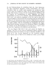
536 JOURNAL OF THE SOCIETY OF COSMETIC CHEMISTS (lO) (11) (12) (13) (14) (15) (16) (17) (18) (19) (20) I. Willis and A.M. Kligman, Aminobenzoic acid and its esters. the quest for more effective sunscreens, Arch. Dermatol., 102,405 (1970). D. J. Cripps and S. Hegedus, Protection factor of sunscreens to monochromatic radiation, Arch. Dermatol., 109, 202 (1974). C. A. Schlagel and E. C. Sanborn, The weights of topical preparations required for total and partial body inunction, J. Invest. Dermatol., 42, 253 (1964). M. A. Pathak, T. B. Fitspatrick and E. Frank, Evaluation of topical agents that prevent sunburn: the superiority of PABA and its esters in ethyl alcohol, N. Engl. J. Med., 280, 1459 (1969). T. M. Macleod and W. Frain-Bell, The study of the efficacy of some agents used for the protection of the skin from exposure to light, J. Dermatol., 84,266 (1971) P. D. Forbes, In the first annual meeting of American Society for Photobiology, Sarasota, Florida, June 10-14, 1973 (Abstract), p 136. S. Zigman, E. Fowler and A. L. Kraus, Blacklight induction of skin tumors in mice, J. Invest. DermatoL, 67,723 (1976). K. G. Hansen, Transmission through skin of ultraviolet and visible radiation 280--500 nm, in "The Bio- logic Effects of Ultraviolet Radiation, with Emphasis on the Skin," F. Urbach, Ed., Pergamon Press, 1969, pp 159-163. M. A. Everett, E. Yeargers, R. M. Sayre and R. L. Olson, Penetration of epidermis by ultraviolet rays, Photochem. and Photobiol., 5, 533 (1966). K. H. Kaidbey and A.M. Kligman, Further studies of photoaugmentation in humans: phototoxic reac- tions, J. Invest. Dermatol., 65,472 (1975). P. Bener, Spectral intensity of natural ultraviolet radiation and its dependence on various parameters, in "The Biologic Effects of Ultraviolet Radiation," F. Urbach, Ed., Pergamon Press, New York, 1969, pp 351-358.

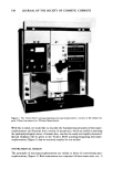
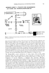
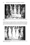
J. Soc. Cosmet. Chem., 29, 537-544 (September 1978) Use of microspectrophotometry in dermatological investigations GARY L. GROVE, ROBERT M. LAVKER and ALBERT M. KLIGMAN Simon Greenberg Foundation, 3401 Market Street, Philadelphia, PA 19104. Received January 25, 1978. Presented at Annual Scientific Meeting, Society of Cosmetic Chemists, December 1977, New York, New York. Synopsis Quantitative estimation of dimensions of structure and amounts of material in skin at the light microscope level has, until now, required time-consuming and tedious methods that are often subject to observer error. We have overcome these problems by using a Vickers M86 scanning-integrating microspectrophotometer. This analytical light microscope detects the amount of light which can pass through a specimen and then electronically converts this value into units of absorbance and projected area. This approach is very versatile and is in fact applicable to any biological structure which can be identified at the light microscope level and in which an appropriate change in color intensity can be realized. The fundamental principles of visible light MICROSPECTROPHOTOMETRY and its application to DERMATOLOGICAL STUDIES that objec- tively evaluate the pathophysiological status of skin are described. INTRODUCTION For many years, light microscopists have been obliged to rely on "eyeballing" to assess such items as acanthosis or atrophy of the epidermis, enlargement or shrinkage of se- baceous glands, and the amount of dermal ground substance in histochemically stained sections. Awareness of the crudeness of such estimates led to the development of more objective methods such as planimetry and stereographic grid analysis. Although these methods are certainly improvements, they also tend to be tedious, time-consuming and prone to human error. These problems led us to consider microspectrophotometry as an alternative method for analyzing dimensions of structure and amounts of material in the samples. Until recently, this technique was considered to be of such an advanced nature that only specialized experts could dream of using it. The advent of commercially available instruments and improved histochemical methods have changed all this. Microspec- trophotometry is currently at the heart of several exciting biomedical research projects and no doubt the next few years will see an increased number of instruments of this type being used in the routine situation for screening, diagnosis and other investigatory purposes. 537
Purchased for the exclusive use of nofirst nolast (unknown)
From: SCC Media Library & Resource Center (library.scconline.org)