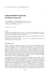
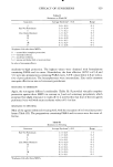
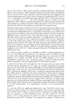
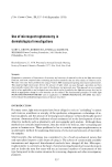
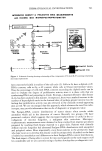
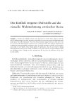
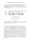
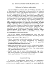
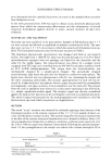
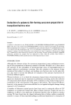
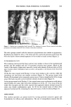
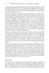
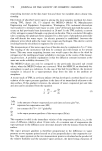
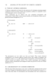
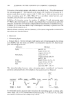
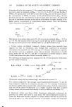
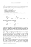
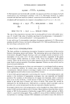
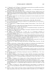
540 JOURNAL OF THE SOCIETY OF COSMETIC CHEMISTS DISTRIBUTIONAL ERROR 80% HETEROGENEOUS OBJECT Equations: m = AB where m,, ma•s of absorbing material in specimen A. absorbance (--log T) B. area of photometeric field T = transmittance MEAN INTEGRATED T A X B = M .301 X I = .301 units T A X B = M !4• .698 x ,25 • .175 .398 x .25 - .100 .222 x .25 = .056 .097 x .25 : .024 • .355 units Figure 3. Distributional error in a model system The specimen is divided into four equal areas of varying chromophore concentrations. The mean value was determined by measuring the transmittance of all areas simultaneously and the integrated value by measuring the transmittance of each area separately. The distributional error increases as specimen heterogeneity increases. At each measuring point the light intensity of the object is transformed into absorbance by a specially designed analog convertor which makes use of the logarithmically decaying voltage of a discharging condensor to transform the signals from the photomultiplier into a train of 10 kHz pulses. Since this circuitry simulates the Beer-Lambert Law, the duration of each of these pulses is proportional to the absorbance at the point. The digitized value of each sample point within the electronically gated measuring field is stored in a computer. At the end of a scanning raster involving over 120,000 measurements of sample area, each small enough to be relatively free of distributional error, these signals are integrated to give a value pro- portional to the amount of absorbing material. Simultaneously a second digital meter gives a reading which is proportional to the area of the specimen which has an absorbance greater than any arbitrary chosen threshold value. By using standards of reference, the absorbance and area meters can be calibrated in absolute units of picograms and square microns, respectively. APPLICATION USING ABSORBANCE MEASUREMENTS Microspectrophotometry was originally designed to estimate the DNA content of an individual cell by measuring the absorbance of Feulgen-stained nuclei. This method has

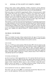
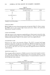
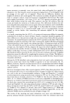
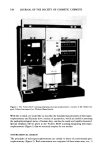
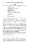
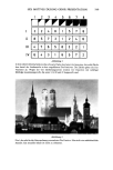
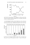
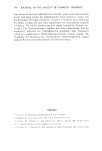
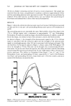
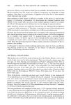
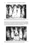
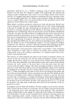
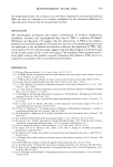
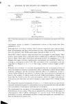
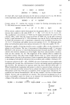
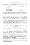
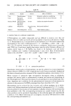
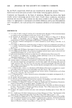
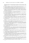
DERMATOLOGICAL INVESTIGATIONS 541 INTEGRATED DENSITY & PROJECTED AREA MEASUREMENTS with ¾1CKERS M85 MICROSPECTROPHOTOMETER monochromltor h] • sc.nner m.sk photomultip specimen SPECIMEN VIEWING SYSTEM MASKING SYSTEM flying-- spot lister scIn photomultiplier ,Integr.ted Density -- signal cted Aree Figure 4. Schematic drawing showing relationship of the components of Vickers M-85 scanning-integrating microspectrophometer been extremely fruitful in studies of the cell cycle (2). Cells in G• have a diploid or 2C DNA contents, cells in G2, a 4C content, while cells in S have intermediate values. Thus the percentage of cells with DNA contents exceeding the diploid mode can be used to evaluate the degree of proliferative activity since it is these cells that are synthesizing DNA and preparing to divide. Psoriasis, a hyperproliferative skin disease (4-6), has been studied in this manner. As expected, there was a marked increase in the number of hyperdiploid nuclei in the lesional skin of perhaps greater interest was the finding that proliferative activity was also elevated in the clinically normal-appearing skin as well. We are encouraged that this approach, which obviates the need for radio- isotopes, may provide information that can be of diagnostic or prognostic value. The Feulgen-DNA content distributions of tumor tissues often reveals subtle anomalies which aid in the detection of cancer. Recently two groups (78) have presented evidence which suggests that microspectrophotometry is useful in the cy- todiagnosis of mycosis fungoides, a malignant skin reticulosis. Microspec- trophotometric measurements were obtained in these studies from imprint specimens prepared by touching fresh biopsy material to a glass slide. Patients with clinically definite mycosis fungoides had abnormal Feulgen-DNA distributions with aneuploid and polyploid values. More importantly, even those patients in the premycotic stage who later went on to develop this disease could be prospectively identified on the basis of subtle but nonetheless real differences in their Feulgen-DNA content distributions. The final impact of being able to screen for mycosis fungoides in the early stages could be considerable and we are currently trying to further develop this cytodiagnostic tool.
Purchased for the exclusive use of nofirst nolast (unknown)
From: SCC Media Library & Resource Center (library.scconline.org)