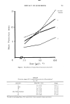
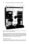
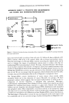
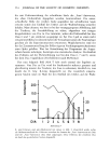
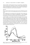
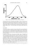
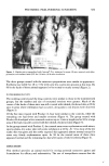
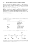
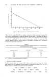
542 JOURNAL OF THE SOCIETY OF COSMETIC CHEMISTS Table I Procedures Amenable to Microspectrophotometric Measurements 1) Histochemical & Cytochemical Staining Reactions: DNA (Feulgen, Gallocyanin-chromalium, methyl green) RNA (Azure B, Pyronin Y) Histones (Alkaline Fast Green, Eosin-Fast Green) Proteins (Naphthol Yellow S, Millon, Sakaguchi) Carbohydrates (PAS) Mucopolysaccharides (Alcian Blue, Mucicarmine, Colloidal iron) 2) Enzymatic Histochemistry: Lysosomal--Bitensky Fragility Test Mitochondrial--monoamine oxidase Pentose Shunt--glucose-6 phosphate dehydrogenase 3) Redox State: Prussian blue of Chevremont-Frederic 4) Natural Pigments: Cytochrome P~450 Hemoglobin 5) Quantitative Autoradiography. Although most absorbance applications have centered on Feulgen-DNA measure- ments, the current surge of interest in microspectrophotometric analysis probably stems from recent development in other histochemical methods. Although the details of these procedures are beyond the scope of this overview, a few examples that are amenable to microspectrophotometric measurements are listed in Table I. In general, these methods enable the detection of tissue chemical changes in the picogram (10 -12 g) range with a routine accuracy of _+ 2%. The use of microspectrophotometry in conjunction with reliable histochemical methods offers several advantages over the conventional form of biochemical analyses ("grind and find"). First, it can be used to make many measurements on minimal amounts of sample tissue. Moreover, multiparameter analyses can often be achieved in the same specimen by using a combination of methods either simultaneously or sequentially. The most important advantage offered by this approach is that it allows the investigator to simply relate observed biochemical changes to the structure of the biological specimen being examined. Thus it is quite possible to measure such things as amount of mucopolysaccharides in the dermis, keratohylin content in the granular layer, the sudanophilia of lipids in sebaceous glands, lysosomal enzyme activity of the basal layer or sulfhydryl or disulfide groups of keratin in situ. With the availability of such instrumentation one no longer needs to be content with making subjective appraisals of staining intensities. Instead it is now quite easy to quantify the precise amounts of specific material of a variety of dermatological specimens from normal or diseased skin. APPLICATIONS USING AREA MEASUREMENTS The Vickers M-85 scanning-integrating microspectrophotometer allows one to measure the projected area of the specimen. We have found this facility to be extremely useful in histogeometric analyses. For example, the need frequently arises to measure the mean epidermal thickness, a parameter which is markedly influenced by disease and experimental manipulations. In the conventional approach, the value

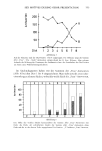
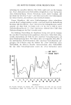
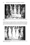
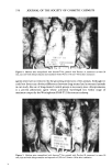
DERMATOLOGICAL INVESTIGATIONS 543 represents the average of 25 to 100 readings at random spots using eyepiece graticule (10). This is not only time consuming and tedious but is subject to considerable errors in specimens with prominent fete-ridge patterns. We are not the first to recognize this problem, as a method based on the Quantiment microdensitometer has been pre- viously reported (11). The key word here is "microdensitometer," because this ap- proach monitors only differences in grey levels--not color intensities. This presents problems when two or more colors are present in the same photometric field and re- quires that the sections be overstained with hematoxylen, which produces a dark blue color, enabling the Quantiment to detect these regions from fainter pink dermal components. With the Vickers M-86 microspectrophotometer these colors can be re- solved and projected area measurements obtained for each colored component. Thus, by measuring the area occupied by the epidermis in a standard size field, one can obtain a global assessment of the dimensions of the viable epidermal compartment with little difficulty. Much of our current research ac.tivity is concerned with developing noninvasive testing procedures for monitoring the physiological status of skin. One extremely promising area is exfoliative cytology, which analyzes cells shed from the body surfaces. Ken McGinley, in our laboratory, has devised a simple detergent-scrub method for quanti- tative sampling and cytomorphological visualization of the cells making up the desqua- mating portion of the horny layer (12). This approach has proved to be very valuable in our studies of psoriasis (13, 14), aging (15), dandruff (16, 17), contact dermatitis (18) and steriod atrophy (19). Many of these studies indicate that changes in corneocyte size permit a sensitive evaluation of altered skin physiology, especially epidermpoieses. Unfortunately, since these cells tend to be quite irregular in shape the techniques that have been employed to date to measure this parameter (axial filar micrometry and polar planimetry) are subject to considerable error. We can, however, rapidly and precisely measure changes in corneocyte size by using the projected area feature of Vickers microspectrophotometry. CONCLUSION The range of applications for both absorbance and projected area measurements covered by this brief survey hopefully has provided some idea of how valuable the microspectrophotometric approach can be. By measuring light absorbance characteris- tics we can analyze dimensions of structure and amounts of material in any biological structure that can be identified at the visible light microscopic level and in which an ap- propriate change in color intensity can be realized. The powerful combination of fast and accuarate geometric and absorbance measurements greatly expands the amount of information which can be obtained from dermatological specimens, viz., scrubs, biopsies, tissue slices, cultures, etc. In fact, the large number of measurements avail- able and the rapidity with which they can be performed means that restraint must be exercised to avoid the unfortunate circumstance of being surrounded by reams of data which have little relevance to the question at hand. REFERENCES (1) R. Barer and F. Smith, Microscope for weighing bits of cells, New Sci., 24, 380 (1972). (2) C. Venderly, Cytophotometry and Histochemistry of the Cell Cycle, in "Cell Cycle and Cancer," R. Baserga, Ed., Marcel-Dekker, New York, New York, 1971, pp 227-268.
Purchased for the exclusive use of nofirst nolast (unknown)
From: SCC Media Library & Resource Center (library.scconline.org)