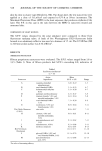
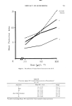
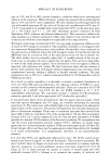
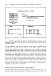
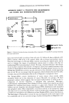
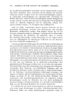
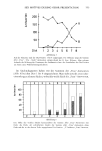
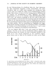
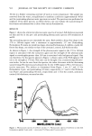
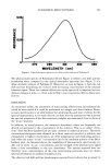
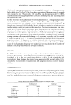
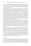
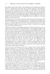
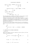
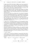
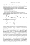
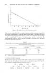
562 JOURNAL OF THE SOCIETY OF COSMETIC CHEMISTS 30 min in a beaker containing one liter of water at room temperature. The sample was removed from the water, reequilibrated to ambient conditions (approximately 48 hr) and the postsoaking photoacoustic spectrum recorded. The previous was performed in triplicate. From these spectra, as will be shown, one can evaluate the sunscreening ef- fectiveness and substantivity to skin of the various formulations. RESULTS Figure 1 shows the ultraviolet photoacoustic spectra of excised, full-thickness neonatal rat skin and the in situ pre- and postsoaking photoacoustic spectra of Formulations A and B. The presoaking spectra are essentially the same. Both exhibit a broad line shape in the 270 to 340-nm region with a maximum at approximately 317 nm. Postsoaking, Formulation B retains its initial line shape, whereas Formulation A exhibits a vastly dif- ferent line shape, one similar to that of the untreated, control, full-thickness skin. As shown in Figure 1, the strength of the photoacoustic signal in the 270 to 340-nm region is associated with the sunscreen agent and the strength of the photoacoustic signal at wavelengths greater than 369 nm is associated with background signal. One can therefore, for the spectra shown in Figure 1, obtain a ratio of the PAS signal at 320 nm to its strength at 370 nm. This ratio can be thought of as a sunscreening effective- ness index. As can be seen from the spectra, the index decreases with the decreasing sunscreening effectiveness and substantivity for the UVB region (290 to 320 nm) pro- tective sunscreen. The indices as determined from Figure 1 are: 8.7 and 2.3 for Formulation A pre- and postsoaking (30 min), respectively 6.7 and 6.4 for Formula- tion B pre- and postsoaking (60 min), respectively and 1.4 for the control, untreated, excised, full-thickness, neonatal rat skin. 0.76 Z O45 o• o•o O. IB & I ! I 2•0 •ZO 5•0 4OO 450 WAVELENGTH (nm) Figure 1. Photoacoustic spectra of newborn rat skin (+). Photoacoustic spectra of newborn rat skin with formulations applied: A pre-soak (-), post-soak (V) B pre-soak (ru), post-soak (O)

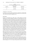
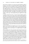
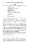
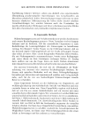
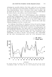
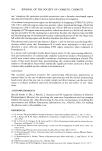
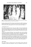
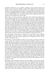
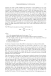
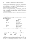
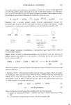
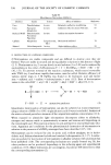
SUNSCREEN EFFECTIVENESS 563 O.5O b.I Z m n., 025 0 I & I , I I 270 i90 $$0 $50 VAVE/ENGTH (nm) Figure 2. Optical absorption spectrum of a dilute alcohol solution of Padimate-O The photoacoustic spectra of Formulation B (see Figure 1) exhibit a red shift and line broadening when compared to the optical absorption spectrum (see Figure 2) of a dilute alcoholic solution of Padimate-O. Moreover, as seen in Figure 1, both the red shift and line broadening are reduced with decreasing concentration of the retained sunscreen agent. These are common effects seen in the spectra of a compound as it un- dergoes changes in state, i.e., from solid to film to less concentrated film to dilute solu- tion (6). DISCUSSION As mentioned earlier, the assessment of sunscreening effectiveness and substantivity would be most useful if it could be performed in a simple and direct fashion. Photo- acoustic spectroscopy is an ideal method for achieving this goal since it allows for the spectral measurement to be made directly on skin thus the parameters which govern the spectral properties of the skin-sunscreen complex are maintained close to those of the "in use" situation. In addition, in actual practice, the measured absorbance values are frequently not directly proportional to the concentration of the solution. That is, so called "devia- tions" from the Beer-Lambert Law are quite common in analytical practice. Therefore conventional absorption data obtained on a dilute sunscreen product in solution, and extrapolated to higher concentration, are often inappropriate and misleading, particu- larly when the actual "in use" concentration lies in the nonlinear portion of the Beer- Lambert plot. As discussed above, the capabilities of photoacoustic spectroscopy en- ß able one to study "in use" concentration, and the strength of the photoacoustic signal bears a close resemblance to the true absorbance. The spectra reported here are therefore more representative of the true sunscreening potentials since we obtain spectral information on undiluted samples in situ. The observed red shift and line broadening as reported here indicate that in actual "in
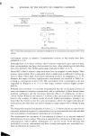
Purchased for the exclusive use of nofirst nolast (unknown)
From: SCC Media Library & Resource Center (library.scconline.org)