J. Soc. Cosmet. Chem., 29, 573-580 (September 1978) In vivo measurement of transepidermal water loss B. IDSON Hoffmann-La Roche, Nutley, NJ 07110. Received September 1, 1977. Presented at Annual Scientific Meeting, Society of Cosmetic Chemists, December 1976, New York, New York. Synopsis An overview is presented of the background and principle methods for MEASURING TRANS- EPIDERMAL WATER LOSS (TWL) IN VIVO. Absolute values of TWL are a function of the particular technique and experimental conditions. TWL will vary with different skin sites and rise markedly if the skin barrier is removed or affected by pathologies. Early gravimetric methods lack sensitivity and require long testing periods as well as large areas of skin. The disadvantages have caused shifts to other techniques where absorption of water vapor is followed by a sensitive physical measurement. The majority of methods are based on determining the increase in moisture content of either a current of dried air or fixed humidity air conducted over the skin. Others have sought to avoid air flow, using changes in conductivity of inorganic crystals. Methods discussed include thermal conductance, electrohygrometry, infrared radiation, electrolysis of absorbed water vapor and calculation of vapor pressure gradient in the layer of air adjacent to the skin sur- face. The mechanism may be an additive effect of neural control of eccrine sweat gland activity and stratum corneum hydration. INTRODUCTION Water exerts a major role in all well-being but particularly in skin health to maintain its desirable soft, flexible mechanical properties (1). The lack of adequate water in the up- per layer of the skin, the stratum corneum, results in dry and chapped skin (2-6). Dermatologic and cosmetic interest has focused on techniques that generate informa- tion on the state and quantity of water in the stratum corneum, the mobility of the water and the influence exerted by components of the stratum corneum on the diffu- sion characteristics of the water (1). Water is lost through skin in two ways, eccrine sweating and transepidermal diffusion. Under severe thermal stress as much as 2 1/hr may be lost as sweat. By contrast, diffu- sional or transepidermal water loss (TWL) is a steady passive process in which water vapor diffuses from the highly hydrated underlying tissues through the avascular stratum corneum, dissolves in it and diffuses to the exterior surface where it evapo- rates. Emphasis is placed on the stratum corneum since this biologically inert membrane--due to its dense, fibrous, lipoprotein matrix--represents the principle physical barrier to the penetration of molecules through the integument (7). The mag- nitude of TWL has been widely used as a measure of the effectiveness of this barrier in dermatological disease states (8-11). In pathologies such as psoriasis, ichthyoses and 573

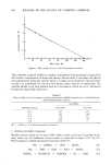
574 JOURNAL OF THE SOCIETY OF COSMETIC CHEMISTS eczema there is impaired barrier function with increased TWL. Normal intact skin has an average TWL of 0.31 mg cm -2 hr -•, rising about ten-fold in psoriasis and eczemas. If the barrier is removed, e.g., by Scotch tape stripping, the TWL rises to 15 to 45 mg a 50-to 150-fold increase over intact skin. No biological membrane of comparable thick- ness offers such resistance (12-14). Water loss through the skin is dependent on environmental factors, of which the most important are the ambient temperature and humidity. Comparison of results of water loss can only be valid if readings are made when these two factors are constant. Like all membrane-diffusion processes, TWL has a characteristic activation energy and therefore its magnitude is temperature dependent (9, 15, 16). Decreasing skin temperature is accompanied by a decreasing TWL. A 5øC fall in skin temperature lowered the TWL by about 45%. The fall appeared to be related to skin temperature and not directly to the reduction in body temperature. A rise of skin temperature of 7 to 8øC doubled the TWL rate (9). A number of"in vitro" and "in vivo" techniques have been described to measure the transepidermal water diffusion from selected areas of the skin. Isolated skin has been studied in vitro in diffusion chambers (17-19). This paper, however, will only be concerned with "in vivo" methodology on human sub- jects. Early techniques of measuring TWL, prior to 1965, have been critically reviewed by Bettley and Grice (8) and Baker and Kligman (20). Analysis of the extensive literature data indicates that the absolute values of TWL largely depend on the technique and experimental conditions used in its measurement (21). However it is evident that with any given technique the values of the TWL consistently vary topographically from skin site to skin site (11, 17, 22). Considerable regional variation was noted in certain areas, even after the readings had been cor- rected for varying horny layer thickness and expressed as diffusion constants. Com- pared with that of the back, the diffusion constant is four times greater on the forehead, nine times greater on the back of the hand and 100 times greater through the palm (20). Technical difficulties have been encountered with all methods. Basically this is because all measurements of TWL must, of necessity, be made under artificial conditions varia- tions in these conditions might be expected to alter the water loss. Broadly, the methods can be classified as "ventilated" and "unventilated": ventilated-•in which a continuous flow of gas or air passes through a capsule attached to the skin and the change in the humidity of the gas is measured by a sensing element in the outflow "unventilated"--in which a container is used with its open end placed on the skin sur- face, the water vapor given off alters the relative humidity within the chamber and this rate of change is a measure of the rate of insensible water loss (11). Any unventilated method is much less satisfactory if the water loss is considerable since water droplets may develop on the skin and fail to evaporate completely. TECHNIQUES Until recently, in vivo determinations have depended upon gravimetric estimation of the water taken up by a hygroscopic medium enclosed in a chamber placed over the skin or removed from a stream of dried air passed through a skin chamber. Pioneering studies (23) involved passing dry oxygen over a small brass chamber attached to the abdomen and collecting the water vapor in the effluent air in freezing coils. In variant
Purchased for the exclusive use of nofirst nolast (unknown)
From: SCC Media Library & Resource Center (library.scconline.org)