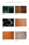
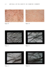
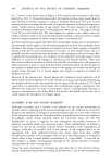
j. Soc. Cosmet. Chem., 34, 407-418 (December 1983) Methods for studying the skin surface L. LOUIS HANTMAN, The Gillette Company, Personal Care Division, Gillette Park, Boston, MA 02106. Received September !983. Presented at the IFSCC/SCCJoint Conference on Skin, San Francisco, September 1983. INTRODUCTION Cosmetic treatments can cause physiochemical and physical changes in the skin surface (stratum comeurn). It is essential for the better understanding of th.e function of skin care products that we should be able to characterize and measure these changes. The use of multiple internal reflection spectroscopy for measuring water content of skin and the deposition of other cosmetic materials on the skin will be reviewed. Color spectrometry or reflectance spectrometry, which is used to study the optical properties of the skin in vivo will also be reviewed. Other methods of examining the skin's surface that will be briefly discussed are Thermography and Multispectral Imaging Analysis. Photographic techniques provide very elegant ways for the visual documentation of the benefits of skin care products. Because they allow the scientist to easily communicate these benefits to technical colleagues and to consumers, they are used extensively in the cosmetic laboratory. Some of these techniques which will be reviewed are low magnification of the skin surface, and high resolution photomicrography using light and electron microscopy. The methods to be reviewed: ß Multiple Internal Reflection Spectrophotometry ß Color Spectrometry ß Thermography ß Multi-Spectral Imaging Analysis ß Macro-Photography ß Light Microscopy ß Scanning Electron Microscopy MULTIPLE INTERNAL REFLECTION SPECTROSCOPY (MIR) The use of Infrared spectroscopy for determining the water content of the skin and testing for the presence of other materials on the skin and the stratum comeurn in 407

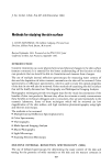
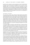
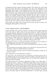
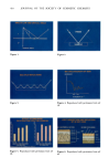
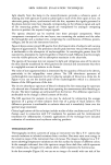
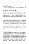
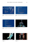
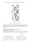
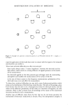
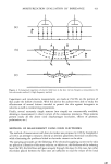
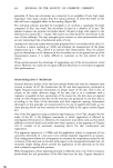
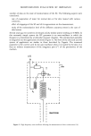
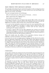
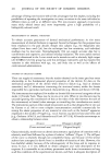
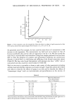
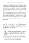
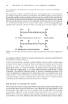
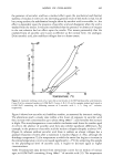
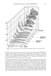
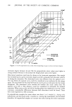
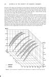
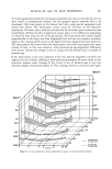
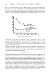
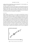
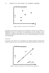
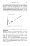
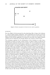
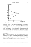
408 JOURNAL OF THE SOCIETY OF COSMETIC CHEMISTS particular has been considered an attractive proposition for some time. Spectroscopic techniques are non-destructive and do not change the state of the substrate. They are particularly useful for moisturization measurements since water has very strong absorption bands in the I.R. region. The use of I.R. spectroscopy for in vivo measurements did not become a possibility until Multiple Internal Reflection (MIR), a spectroscopical technique, had been developed. Incidently, this technique has also been variously described as Attenuated Total Reflectance (ATR) and Frustrated Multiple Internal Reflection (FMIR). MIR was originally developed for the measurement of deposits on surfaces and has been applied in recent years for the determination of materials in the stratum corneum. THE PRINCIPLE OF THE METHOD The method takes advantage of the well-known optical principle of total reflection that follows from Snell's Law of Refraction (Figure 1). When a light beam passes from a medium of high refractive index to a rarer medium, at the critical angle of incidence the light will just graze the surface. At smaller angles of incidence, the light is refracted into the rarer medium at greater angles of incidence, the light is totally reflected back. On internal reflection, all the energy is reflected however, the beam appears to penetrate slightly beyond the reflecting surface before returning. If a material which can absorb radiation is placed against the reflecting surface (Figure 2), the reflected light will be attenuated by the absorption of this energy. In the case of skin, which contains absorbing chromophores, for example, water, then the attenuation of the reflected light beam will be proportional to the number of absorbing chromophores that the light encounters in its path. The attenuated radiation is measured by a spectrophotometer. The spectrum obtained by plotting the MIR versus wavelength can be very similar to that obtained with transmission spectroscopy. Because the absorption band obtained from a single reflection is rather weak, Harrick (1) further modified the original technique [which had been developed by Fahrenfort (2) in the early 60's] by multiplying the amount of absorption with a truncated (trapezoid) prism which increased the number of reflections (Figure 3). If we have a spectrophotometer specially equipped with an MIR prism and the skin is placed against the prism, in principle, the degree of attenuation can be used for obtaining an IR spectrum of the skin. Originally, spectra for a whole range of household and cosmetic products and their ingredients were analyzed by applying the material to the prism, as described by Puttnam (3) et al. By using a V-shaped prism, which was able to accommodate the edge of the hand (the hypothenic eminence), it became possible to study retention by the skin of components present in products applied to the skin. Gloor (4) et al. carried out a detailed study on skin hydration by ATR-IR, following up on earlier techniques discussed by Puttnam and Baxter and by Osberghaus (5). Instead of measuring the water absorption band directly, these workers found it more useful to determine changes in the intensities of the protein absorption bands (Figure 4). The intensity of the polypeptide Amide I band at 1645 cm -• depended on the amount of water present in the stratum corneum. On the other hand, the intensity of the polypeptide Amide II band at 1545 cm -• was insensitive to water content and could, therefore, be used as a calibration standard. Using this technique, Gloor et al. were able
Purchased for the exclusive use of nofirst nolast (unknown)
From: SCC Media Library & Resource Center (library.scconline.org)