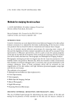
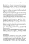
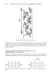
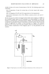
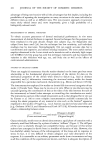
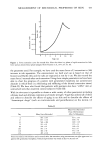
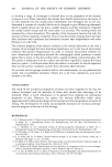
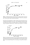
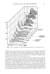
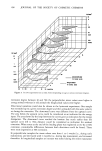
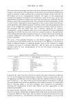
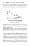
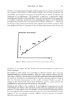
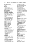
MEASUREMENT OF MECHANICAL PROPERTIES OF SKIN 433 0.4 g 0 2 4 6 8 10 12 Time (s) Figure 1. Force extension curve for normal skin. Note that there is a phase of rapid extension for little force and an almost linear phase (adapted from Gunner et al., 1977, ref. no. 10). the parameter used. For example, we have used the mean force of 3 extensions to 30% increase in tab separation. The extensometer we built and use is based on that of Gunner and Burlin (10), and the tabs are separated at rest by 1 cm. We also record the mean force 5 seconds after each extension. Using these simple parameters we have been able to chart the progress of a patient with generalized morphoea (an autoimmune disorder in which there is a thickening and stiffening of the dermal connective tissue) (Table II). We have also found that patients with psoriasis also have "stiffer" skin at uninvolved sites than matched control subjects (Table III). With in vitro tests it is possible to obtain a wide variety of other parameters including ultimate load and ultimate extension and tensile strength. Vogel has utilized all of these and others to describe the effects of aging in rat skin and to describe the effects of "desmotropic drugs" (such as corcicosteroids and penicillamine) on the dermis. (2) Table II Day Mean Force Recorded Mean Force Recorded From 30% Extension After 5s. Relaxation 0 593 ñ 195 437 ñ 155 17 875 ñ 139 658 ñ 76 42 540 ñ 206 340 ñ 118 75 213 ñ 41 155 ñ 13 89 210 ñ 61 148 ñ 24 103 553 ñ 75 433 ñ 57 110 492 ñ 30 472 ñ 30 154 285 ñ 139 158 ñ 53.9 Forces recorded at 3% extension and 5 seconds after 30% extension using extensometer on right upper arm in a patient with generalized morphoea. Clinical improvement was noted at days 75, 89, and 154.

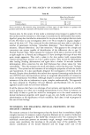
434 JOURNAL OF THE SOCIETY OF COSMETIC CHEMISTS Table III Mean Force Recorded Mean Age From 30% Extension Psoriatics 37.7 -+ 22 449 __ 149 Control subjects 31.8 _+ 14 309 -+ 107 Forces recorded at 30% extension using extensometer on flexor aspect of right forearm in 9 patients with psoriasis (uninvolved skin examined) and 8 matched normal control subjects. Torsion tests. In this variety of test mode a rotational stress (torque) is applied to the skin surface and the resistance to the torque is recorded as the deformation that results. Agache's group has described an instrument for in vivo use that employs this test mode (15,16). The force in one investigation (28.6 x 10⁻³N.m) resulted in angular displace- ment of the skin 2-6 ø. They corrected for skin thickness and were able to calculate a number of parameters including "immediate distension," "final distension" (after 2 minutes), "delayed distension," and "late retraction." This appears to be a simple and convenient test system capable of identification of the differences due to age. (3) Normal Traction Tests. These measure the resistance of the skin to traction on the skin at right angles to it. Pierard and colleagues (17) have developed this type of test with quite simple apparatus. They stick a plate to the skin surface (with cyanoacrylate cement) and produce traction on it by a pulley system. They record the deformation after loading (loading deformation) and again after a further 20 seconds (residual deformation). After correction for skin fold thickness they obtain values for "skin extensibility." They also measure the recoil and obtain a value that they term "biologic elasticity." The system has the limitation of recording the properties of both the dermis and hypodermis. It is also specifically designed for investigation of the skin of the forearm. Despite these drawbacks the authors have reported interesting results from the use of PUVA and corticosteroids as well as in congenital abnormalities of connective tissue. (4) Indentation Test Mode. Although simple in concept the measurement of the resistance of the skin to indentation is quite complex mathematically. This approach has been adopted by Vlasblom (18) and Dikstein and Hartshtark (19). As yet this approach cannot claim to have been validated. It will be obvious that there is no shortage of test systems, and standards are available to give guidance as to their use in making measurements in vivo. There is, however, no firm body of data to guide the would-be investigator as to the usual variations in dermal mechanical properties with regard to age, sex, site, bodily activity, and drug administration or the significance of such variability. Such data is vital to progress in this area of skin biology. TECHNIQUES FOR MEASURING PHYSICAL PROPERTIES OF THE STRATUM CORNEUM The stratum comeurn is less hererogenous than the dermis but is nonetheless a highly complex structure. It contains a system of protein filaments set in a matrix of unknown composition and packaged in flat shield-like desicated cells--the corneocytes. The individual corneocytes possess an extremely tough, strongly cross-linked protein- aceous outer membrane that develops in the granular layer of the epidermis. The relative contribution of the keratin filaments and the tough outer coat of the
Purchased for the exclusive use of nofirst nolast (unknown)
From: SCC Media Library & Resource Center (library.scconline.org)