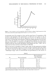
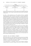
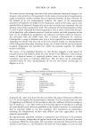
MEASUREMENT OF MECHANICAL PROPERTIES OF SKIN 431 Table I In vivo and in vitro Skin Thickness Determination by Ultrasound Ultrasound Skin Thickness (mm) Subject In vivo In vitro 1 0.91 1.52 2 o.87 1.19 3 1.06 1.52 4 o.68 1 .o6 5 o.68 1.14 6 1.06 1.37 7 1.14 1.52 Mean 0.91 1.33 +s.d. +0.18 _+0.20 TECHNIQUES FOR MEASUREMENT OF MECHANICAL PROPERTIES OF THE DERMIS Above I have given reasons for the comparatively low interest in bioengineering techniques from skin scientists. But it should be emphasized that it is possible to obtain highly relevant information from mechanical testing from at least two parts of the skin, the dermis and the stratum corneum. The dermis is a complex meshwork of collagen, reticulin and elastic fibers set in a gel-like matrix with a high content of glycosaminoglycan and proteoglycan. Through this beterogenous structure course blood vessels and nerves, and it is pierced by hair follicles and sweat ducts. It is firmly bonded to the epidermis above and joined (though somewhat less firmly) to the subcutaneous fat beneath. As already discussed, the dermis in vivo is in a state of resting tension that has a particular orientation to the body axis at different sites and differs in properties and functions according to the precise body site examined. To make any sense of an investigation of derreal mechanical properties all these issues must be kept in mind. STANDARDIZATION A major problem for the subject has been the lack of comparability of the work from different laboratories. Individual investigators have used different instruments and/or employed widely varying test procedures, completely vitiating any comparison between the results. To surmount this difficulty the standardization subcommittee of the International Society of Bioengineering and the Skin has taken on the task of publishing standards after extensive discussion and consultation. Discussion docu- ments (1) and a suggested standard (2) for the in vivo testing of derreal mechanical properties now exist. It should now be possible to compare results obtained in Cardiff with those obtained in San Francisco. PARTICULAR TECHNIQUES AND INSTRUMENTATION FOR INVESTIGATING THE DERMIS The dermis is an excellent example of a structure where it may be helpful to perform both in vivo and in vitro tests. Investigations performed in vivo have the supreme

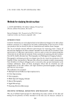
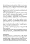
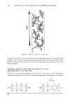
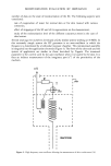
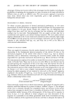
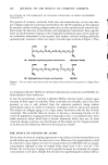
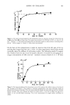
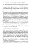
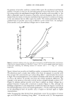
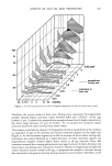
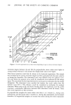
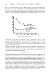
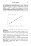
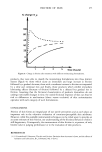
432 JOURNAL OF THE SOCIETY OF COSMETIC CHEMISTS advantage of being non-invasive with all the advantages that this implies, including the possibilities of repeating the investigation on many occasions in the same individual at different times as well as at different sites. The non-invasive approach circumvents many tricky ethical issues and, more importantly, gives a high probability of a biologically relevant result. MEASUREMENT OF DERMAL THICKNESS To obtain accurate parameters of dermal mechanical performance in vivo some measurement of derrnal thickness is required. Several techniques for this purpose have been employed in the past decade. Simple skin calipers (e.g., the Harpenden skin caliper) have been used (3,4), but the technique has low sensitivity, and individual readings may be inaccurate. Xeroradiography (5,6) can supply accurate data but is cumbersome and expensive, and utilizes ionizing irradiation. The most useful method employs ultrasound in the A scan mode and a transducer with a relatively high output (20-30 MHz) (7,8). Our group has used this technique extensively and has reported the variation in skin thickness with age, sex, and body site as well as the effects of corticosteroid administration. STUDIES ON ISOLATED DERMIS These can supply (a) reassurance that the results obtained on the intact part bear some relationship to the fundamental physical properties of the dermis (b) data on the mechanical properties of the dermis when tested to failure (e.g., load at ultimate extension) and (c) information concerning the structural moiety within the dermis responsible for a particular mechanical characteristic [e.g., Minns and Steven 1979 (9)]. The instrumentation employed for studies on dermis falls into several categories of test mode: (1) Tensile Tests. These may be in vivo or in vitro. When in vivo the tests may be uniaxial (ignoring the contribution of skin at the sides of the skin between the tabs of the instruments) or biaxial (when attempts at controlling this contribution are made). The instruments used are either simple tensometers such as that designed for in vivo use by Gunner et al. (10) or more complex instrumentation available commercially for testing the elastic properties of any material in vitro such as the Instron © apparatus employed in the studies of Vogel (e.g., 11,12). Suction cup devices have also been used [e.g., Grahame 1970 (13), and Alexander and Cook 1977 (14)]. All tensile tests attempt to obtain a relationship between stress (Force) (Change in length) and strain (Cross sectional area) (Original length) Characteristically, tensile tests on dermis demonstrate a rapid phase of extension with a relatively small load followed by a phase of relative "stiffness" (Figure 1). The stiff part of the curve is relatively (though not completely) linear, and because of this several authors have attempted to derive from it a modulus of elasticity of skin. However, this has doubtful validity because of the inhomogeneity of the tissue and its response to stress. In fact, it is very difficult to obtain adequate and valid descriptors of the response. It is reasonable to use arbitary parameters under these circumstances as long as all the conditions of the experiment are known, and there is adequate description of
Purchased for the exclusive use of nofirst nolast (unknown)
From: SCC Media Library & Resource Center (library.scconline.org)