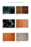
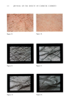
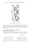
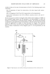
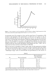
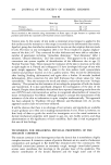
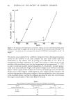
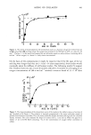
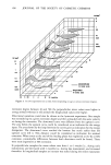
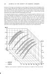
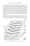
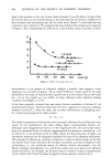
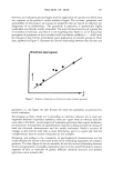
MEASUREMENT OF MECHANICAL PROPERTIES OF SKIN 435 corneocytes to the overall strength of the horny layer is unknown. The links between individual corneocytes are clearly also of importance to the mechanical stability of the horny layer and are of several varieties. The desmosomal connections between epidermal cells remain within the stratum corneum, but whether they have a functional importance at this site is unknown. The corneocytes interdigitate with each other through a series of foldings in the cell membrane, and this could be one of the major ways of maintaining the strength and stability of the overall structure. Between the corneocytes is a material which appears to be lipid in nature and may have great importance for desquamation. This too must contribute to the mechanical properties of the horny membrane. (1) In 17itro Test Procedures. Isolation of the stratum corneum is no easy task. Cantharidin blister roofs have been used, but strips obtained after some form of dermo-epidermal separation and further maneuvers to remove epidermal cells are the most usual substrates for mechanical tests. Portions of heel callus have also been employed, but the doubt remains as to whether the results recorded bear any relationship to the properties of stratum corneum of other sites. Clearly, the act of obtaining the stratum corneum can in itself alter its integrity and influence the results of mechanical studies. An even more severe constraint concerns the avidity of the horny layer for water and the dependence of its mechanical properties on its water content. Tests in vitro must be performed in controlled conditions in which the ambient temperature and humidity are known and variable. Most information has been obtained from tensile tests similar to those performed on the dermis in vitro and performed with similar if not identical instruments (e.g., 20,21). Other types of mechanical testing procedures, performed on the stratum corneum in vitro previously have included thermomechanical analysis [Humphries and Wildnauer 1971 (22)], and indentation characteristics using a rapidly indenting needle to record resisting forces to penetration [Guibarra et al. 1981 (23)]. (2) In 17ivo Test Procedures. Few reliable tests of the mechanical properties of the stratum corneum in situ have been described. There is a desperate need for such tests to assist the development of drugs that modulate keratinization and desquamation. Unfortunately, there are major intrinsic difficulties to the development of such tests which surpass even those associated with in vivo dermal tests. As mentioned previously, the stratum corneum is quite hygroscopic, and ideally an environmental chamber should be used for these experiments. However, as long as the ambient temperature and relative humidity are known, constant, and in the "comfort range" (temperature 17-21øC and relative humidity 50-65%), this is sufficient for most purposes. Because of the tight fixation of the stratum corneum to the epidermis that forms it and its relative hardness, it is quite difficult to devise tests that measure its properties alone. Despite all these difficulties some tests have been developed. Christensen et al., 1977 (24), described a dynamic test using an electrodynamometer employing a gas suspended armature which they claimed supplied information on the viscoelastic properties of the stratum corneum. A force of approximately 5 Gms. was applied and the instrument was set to cycle at 1-2 cycles per second. From the major slope of the hysteresis loop produced it was possible to calculate the "dynamic spring rate" which is analogous to Young's Modulus of elasticity. Changes after application of emollients to the skin were described, but there appear to be no other validating experiments. As our group has major interests in the process of keratinization we have made special efforts

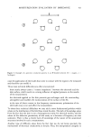
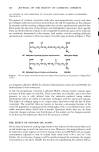
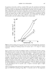
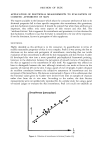
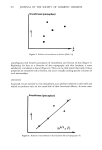
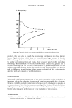
436 JOURNAL OF THE SOCIETY OF COSMETIC CHEMISTS to develop a range of techniques to furnish data on the properties of the stratum corneum in vivo. Point indentation has already been briefly mentioned in discussion of in vitro methods, but the needle point identometer was developed for in vivo use. Essentially it consists of a needle (which can be changed to give different tip diameters) which is rapidly driven into the skin a variable distance and then withdrawn. Typically the needle travels 10/xm and the cycle takes 4 m seconds. The resisting forces are measured by a force transducer. The rapidity of the movement ensures that only the stratum corneum responds. Using this device it was found that resisting forces decrease after hydration with emollients and transiently increase after delipidization with ether [Henley et al., 1981 (25)]. The cohesive property of the stratum corneum in the vertical dimension is not only a measure of its strength but has a functional significance as it is the loss of intracorneal cohesion that permits desquamation. In order to measure intracorneal cohesion we have constructed an instrument (termed the cohesograph) which possesses a central piston that is struck to the skin surface with a rapidly bonding cyanocrylate glue (26). The piston is withdrawn from the surface and the force required to rupture the horny layer (at a point 2-3 cell layers down from the surface) is recorded. As may be expected, there are site and sex variations, and the force decreases after hydration. As yet there are few groups involved with in vivo measurement, no one preferred test mode, and no published standards. Clearly this is far from satisfactory, and much remains to be done. CONCLUSION The study of the mechanical properties of tissues has been impeded by the way the subject developed and the attitudes of those who should take advantage of its potential. There is much information to be gained from studying the mechanical properties of skin. However, the subject has intrinsic difficulties which unless appreciated will confound the design of test systems and the interpretation of results of testing. The development of criteria and acceptable standards are necessary prerequi- sites for progress in this area of skin biology. REFERENCES (1) J. C. Barbenel and P. A. Payne, In vivo mechanical testing of dermal properties (discussion document), in Report No. 1, International Society for Bioengineering and the Skin, Sub-Committee for Standardisation, 1982. (2) J. C. Barbenel, In vivo mechanical testing of dermal properties (Standards), in Report No. 1, International Society for Bioengineering and the Skin, Sub-Committee for Standardisation, 1982. (3) B. McConkey, G. M. Fraser, A. S. Bligh and H. Whiteley, Transparent skin and osteoporosis, Lancet, 693-695 (1963). (4) J. D. Kirby and D. D. Munro, Steroid induced atrophy in an animal and human model, Br. J. Dermatol., 94, Suppl. 12, 111-119 (1976). (5) R. Marks, P.J. Dykes and E. Roberts, The measurement of corticosteroid induced dermal atrophy by a radiological method, Arch. Dermatol. Res., 253, 93-95 (1975). (6) P.J. Dykes and R. Marks, Measurement of skin thickness: A comparison of two in vivo techniques with a conventional histometric method,J. Invest. DermatoL, 69, 275-278 (1977). (7) C. Y. Tan, R. Marks and P. A. Payne, Comparison of xeroradiographic and ultrasound detection of corticosteroid induced dermal thinning,J. Invest. DermatoL, 76, 126-128 (1981).
Purchased for the exclusive use of nofirst nolast (unknown)
From: SCC Media Library & Resource Center (library.scconline.org)