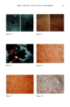
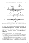
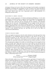
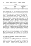
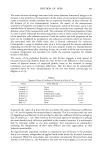
446 JOURNAL OF THE SOCIETY OF COSMETIC CHEMISTS Table II The Stabilizing Effect of Ascorbic Acid on Some Mechanical Properties of Rat Tail Tendon from Animals of Different Ages Age of Animal Change Change (months) in Extension Modulus (%) in Stress at Break (%) 1.5 71 118 6.5 57 75 8.6 59 66 24.0 32 15 Results derived from stress/strain curves of rat tail tendon extended to break in 0.15M NaCI containing 20mg ascorbic acid 100 ml •, at 36øC. The changes in extension modulus and stress are relative to values obtained in 0.15M NaCl at 36øC. was 36øC, i.e., body temperature for rat. The ascorbic acid causes a remarkable strengthening of the tendon, and though the concentration used was much higher than normal physiological level, even this level (2mg 100 ml 1) produces about half the effect shown in the table. The changes due to ascorbic acid become less with age, and a speculative interpretation of this observation is that ascorbic acid behaves as an easily disrupted cross-link (except for the small percentage of cases we have observed with very young tendon in which the strengthening is irreversible). How this could be is not known perhaps ascorbic acid may form compounds with the existing reducible cross-links resulting in the kind of multi-functional links suggested in the previous section. Some support is given to this speculation by the following result: tendon which had been transformed to the rubbery state was unaffected by the presence of ascorbic acid in stress/strain experiments. That is, the native collagen conformation is required for a reaction to occur with ascorbic acid. Whatever the mechanism underlying the capacity of ascorbic acid to stabilize collagen tissues, the behavior is consistent in particular situations. Gould (20), in summing up studies concerning ascorbic acid and collagen, concludes "an analysis of all the data available suggests that ascorbic acid may be involved in the maintenance of rapidly formed labile collagen but is probably not involved to the same degree in the maintenance of most types of structural collagen." In the light of the results just presented, this statement may be interpreted as saying that ascorbic acid is essential to stabilize newly-synthesized tissues where mechanical strength and thermal stability is important, e.g., wound healing of all kinds. Once the normal maturation processes are under way the maintenance becomes less important, and as our results indicate, the effect of ascorbic acid decreases with age. INORGANIC IONS AND THERMAL STABILITY We have just discussed two of the components of the natural environment of collagen fibers (namely oxygen and ascorbic acid) which markedly affect their properties. The inorganic ions in physiological solution mentioned earlier are also part of the environment, but their function with respect to collagen is not clear (although of course they are essential to the functioning of the cell). A good approximation to •'physiological" solution has the following composition, expressed as grams of neutral salt in 1000ml of water: NaC1, 9 KC1, 0.4 CaC126H20,

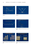
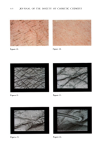
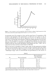
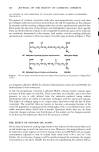
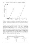
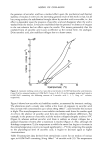
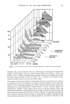
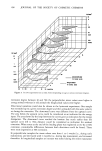
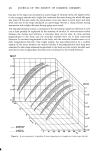
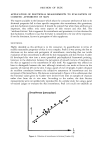
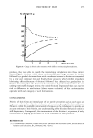
AGING OF COLLAGEN 447 0.25 NaHCO3, 0.2 (23). In large concentrations, these salts cause changes in the melting point of both collagen molecules in dilute solution, and collagen fibers, but at the in vivo concentration given above they are ineffective (24). Thus NaC1, which is by far the most abundant of the salts in tissues, decreases the melting point of collagen by only --•0.25øC compared with pure water. The relative proportion of the ions could be important in in vivo aging. Preliminary experiments in vitro (Figure 7) show how the isometric melting curve responds to different salt combinations after six months storage followed by testing in O.15M NaC1 (25). All the salts were present at the normal in vivo concentration. The addition of KC1 or CaC12 to NaC1 causes a greater increase in thermal stability than occurs in NaC1 alone. With all the salts present, the collagen stability is intermediate between that achieved in NaC1 alone and NaC1 with KC1 or CaCI 2 added. Because C1- is massively abundant in all the combinations, K + and Ca ++ ions apparently have a marked influence on thermal stability. These two ions, K + and Ca ++, have now been shown to also greatly affect the stress/strain properties of both tendon and sheep skin at body temperature (25). In attempting to understand phenomena such as these just described it is useful to realize that collagen and its attendant glycosaminoglycans can be viewed as hydrated polyanionic aggregates, and that fluctuations in the concentration of cations such as K + and Ca ++ could well alter stabilizing interactions between macromolecules. Comments on other interrelations between collagen and glycosaminoglycan will be made later. COLLAGEN TYPES The preceding discussion centered on the collagen present in tendon. It is now established that collagen is actually the family name of a group of genetically distinct molecules in which at least four types are clearly recognized (26). Type I collagen, which constitutes approximately 90% of human body collagen, occurs in skin, tendon, bone, ligament, fascia, dentin, and blood vessels. Of the remaining 10% of body collagen, type II occurs predominantly in cartilage and nucleus pulposus, type III is distributed as for type I, except in bone and tendon, and type IV is involved in basement membranes, for example, kidney glomeruli. The essential differences between these various types is in the amino-acid sequence of the three chains which make up the triple-helical molecule and whether the three chains are identical in a given molecular type. Thus, a type I collagen molecule contains two chains which are identical and a third which is different, whereas type III collagen has three identical chains. All these genetic types of vertebrate collagens exhibit the aidehyde cross- linking system outlined earlier, so that while the earlier discussion has been concerned with tendon, the arguments should apply to the collagen of other tissues, e.g., skin. GLYCOSAMINOGLYCANS AND COLLAGEN FIBRILS The various members of the family glycosaminoglycan are in general associated with different collagen types and are now considered to be involved in the control of collagen fibril size, among other functions (27). Thus, type III collagen has finer fibrils
Purchased for the exclusive use of nofirst nolast (unknown)
From: SCC Media Library & Resource Center (library.scconline.org)