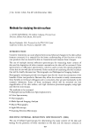
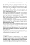
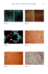
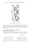
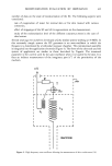
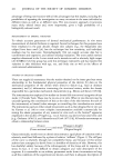
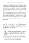
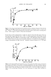
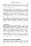
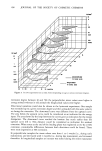
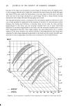
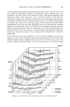
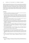
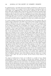
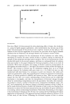
SKIN SURFACE EVALUATION TECHNIQUES 413 ATI•NUATOR ROTATING DISC VIEWING APERTU•,BE R SKIN r LG (INVERSE REFLECTANCE) 0.8 PHOTODE'I•CTOR 0.7 • : , ,. AMPIJFI•R 0.8 p • i•r--• 0.6 f • --• I:1 C1RON• 0.4 O. • , • I I 450 600 650 600 6S0 700 WAVELENGTH (NM) Figure 7. Reproduced with permission from ref (6) Figure 8. Reproduced with permission from ref (6). LG (INVERSE REFLECTANCE) 0.4* [- 450 600 550 600 650 700 WAVELENGTH (NM) Figure 9. Reproduced with permission from ref (6). Figure 10 Figure 11. Figure 12.

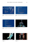
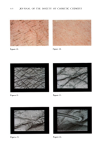
414 JOURNAL OF THE SOCIETY OF COSMETIC CHEMISTS scattered. The longer the wavelength the deeper the penetration. Dent determined that violet-blue light (300-450 nm) does not penetrate beyond the stratum granulosum while the red wavelengths (480 to 800 nm) are able to penetrate further into the dermis. Therefore, for good detailed texture of skin surface, you should use the violet-blue light. The green-red light shows the veins and blood vessels but gives no surface detail. Quattrone and Laden (9) in 1975 confirmed this work and used low magnification photographs to characterize the degree of dryness of dry skin. A panel of judges was used to examine a series of photographs of skin with varying degrees of dryness. They assigned scores according to the severity of flaking. This technique was used to evaluate the efficacy of emollient creams. In Figures 13 and 14, we can see some typical photographs comparing the same site before and after a moisturizing treatment, first, (Figure 13) as it appears in black and white and, next, (Figure 14) another study using color photography. LIGHT MICROSCOPY Another technique for the evaluation of the effects of cosmetic treatments on the skin is the use of the light microscope. In our laboratory, we use a Wild-Heerbrugg Stereomicroscope with fiber optics and a flash attachment. The magnification we get is between six and one hundred times, but typically we operate at about twelve times. Figure 15 shows a typical set up. In Figures 16 and 17 are shown the effects of a moisturizing treatment on dry skin. In another study, we used this technique to demonstrate skin soiling and cleaning. In this case, the skin was soiled with a mixture of carbon and mineral oil (Figure 18). In Figure 19 we see the improvement when the skin was washed with soap and water however, some of the soil is left on the skin. A cosmetic scrub seemed to do a better job of removing the soil (Figure 20). SCANNING ELECTRON MICROSCOPY With the Scanning Electron Microscope (SEM), the effects of topically applied materials on the skin can be assessed at a much greater magnification and depth of field than with light microscopy. With our AMR 1200B model, we can g•t magnifications over 50,000, although there is little practical value over 30,000. For the SEM, we use replicas of the skin rather than use the skin itself. This is done by casting a negative impression of the skin. We use a silastic elastomer for this and then follow with a positive replica of epoxy or polyethylene. The value of using replicas for comparing treatments is that the same site can be used before and after treatment. If repeated replicas may alter or remove some surface characteristic such as desquamous material from dry skin, adjacent sites may be used. In Figures 21 through 25, the appearance of the skin can be studied as magnification is increased, starting at 50, then 100, 200, 500, and 1,000 magnifications. Valleys and ridges of the skin pattern can be seen, also veilus hair with cuticle. In the next series (Figures 26 and 27), the effects of moisturization on dry skin can be seen. In the case of dry skin, fissuring between areas of apparently adherent corneocytes can be seen as well as scale sloughing from the epidermal surface. The hydrated skin, on the
Purchased for the exclusive use of nofirst nolast (unknown)
From: SCC Media Library & Resource Center (library.scconline.org)