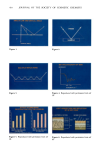
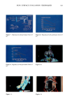
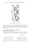
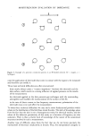
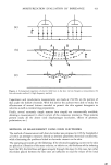
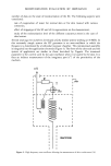
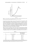
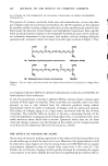
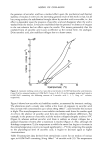
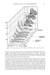
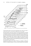
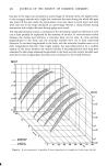
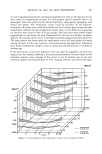
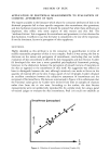
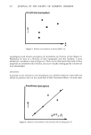
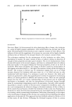
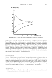
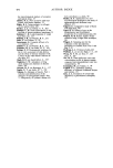
SKIN SURFACE EVALUATION TECHNIQUES 409 to determine the water content of stratum corneum. They carried out a study using both oil-in-water and water-in-oil emulsions. The ratio of the absorption of the polypeptide Amide I band to the polypeptide Amide II band they referred to as the moisturizing factor (Figure 5). For untreated skin, this was in the region of 1.5 and increased to about 1.8 to 2.0 after the skin was moisturized with the emulsions. In this figure, we have bar graphs which plot the moisturizing factor for both oil-in-water and water-in-oil emulsion treatments before treatment and then after 10 minutes, 20 minutes, and 30 minutes of treatment. There was no significant difference between the oil-in-water and water-in-oil emulsions' moisturizing factors. As can be seen, both treatments were quite effective after 10 minutes of treatment, but applications for longer periods did not appear to provide additional moisturizing benefits. I have studied quite a large number of papers for this review, and I have included in the bibliography those papers that I have used. COLOR (REFLECTANCE) SPECTROMETRY Another challenge for the cosmetic scientist is to be able to examine optical properties of the skin, such as surface color, in vivo and attempt to correlate visual and instrumental methods. As pointed out by J.B. Dawson (6) et al., surface color may be quantified by using the CIE system of tristimulus values or by measuring its reflectance spectrum. They make the point that the former method is valuable for color matching, but the latter technique gives more information about the substance generating the color. Dawson et al. suggested an approach based on a parameter which is the logarithm to base ten of the inverse of reflectance (abbreviated to L.I.R.). This approach is based on a mathematical treatment of the factors which contribute to the visual appearance of the skin and involves three assumptions: 1. Skin consists of several layers, each of which homogeneously transmits and scatters light. 2. The transmission and scattering of light can be described by formulas which were developed to explain the diffuse reflectance of powders. 3. The interface between the layers does not modify the transmittance of light by the system as a whole. These assumptions may be demonstrated in Figure 6. The horny layer in the anatomical section corresponds to the fibrous protein layer in the simplified model, the second layer contains the melanin, the blood vessels in the dermis are represented by the hemoglobin layer, and the fourth layer represents the subcutaneous fat and collagen. The essential parts of the instrument, represented in Figure 7, are a light source to illuminate the surface, a means of collecting the light reflected from the surface, a spectral analyser or monochromator, and a means of measuring the light intensity and computing the desired function. The system uses fiber optics to carry the light from the source to the surface and the reflected light to the spectral analyser. Specular reflection from the surface was avoided by mounting the fiber optics in a metal cowl at an angle of 45 ø to the skin surface. A dual channel system is used in order that two adjacent areas of the surface might be examined simultaneously. A third fiber optic, carrying

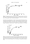
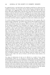
410 JOITRNAL OF THE SOCIETY OF COSMETIC CHEMISTS SNELL'S LAW AND CRITICAL ANGLE CRITICAL ANGLE Figure 1. Figure 2. MULTIPLE REFLECTIONS Figure 3. MIR SPECTROGRAPH OF SKIN ABSORBANCE 1645 1545 WAVELENGTH, cm-1 Figure 4. Reproduced with permission from ref (4). EFFECT OF EMULSIONS ON HYDRATION STRATUM CORNEUM MF MF 20 20 19 19 17 I 17 1õ 16 1S 1 14 I 14 13 13 12 12 11 11 10 -- 10 BEFORE 10 20 30 BEFORE 10 20 30 MINUTES AFTER TREATMENT MINUTES AFTER TREATMENT O/W EMULSION W/O EMULSION FROM M. GLOOR ETAL ARCH DERMATOL CAS (1981} 271: Figure 5. Reproduced with permission from ref (4). LIGHT ABSORPTION AND REFLECTION BY IN VIVO SKIN A} ANATOMICAL SECTION (B} SIMPLIFIED MODEL Figure 6. Reproduced with permission from ref (6).
Purchased for the exclusive use of nofirst nolast (unknown)
From: SCC Media Library & Resource Center (library.scconline.org)