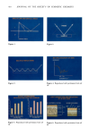
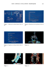
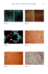
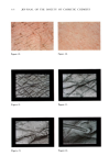
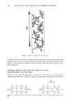
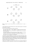
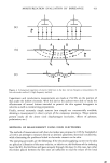
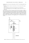
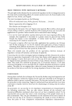
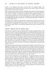
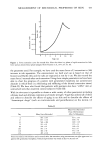
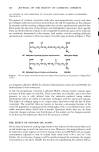
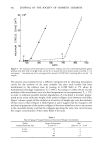
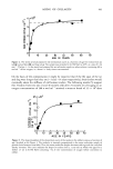
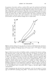
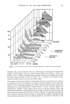
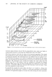
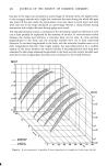
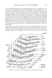
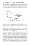
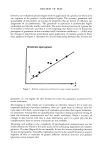
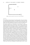
SKIN SURFACE EVALUATION TECHNIQUES 411 light directly from the lamp to the monochromator, provides a reference point. A rotating disc with apertures is used to admit light to each of the fiber optics in turn. An electronic gating device, synchronized with the disc, separates the signals generated in the photo detector into three channels corresponding to the reference signal and each of the measuring probes. These signals are manipulated by analogue computing devices to produce the required output signals. The spectra obtained can be resolved into three principal components. These components correspond to the two layers, one containing the melanin and the other the hemoglobin and a residual term, comprising the contributions of fibrous protein, collagen, and fat and the instrument function. Figure 8 shows some typical LIR spectra from the forearm skin of subjects with varying degrees of pigmentation. The prominent double peak between 510 and 600 nanometers is attributable to the absorption of oxyhemoglobin. At wavelengths greater than 620 nanometers, absorption by oxyhemoglobin is relatively small. Melanin absorbs strongly over the visible spectrum, with its absorption maximum in the ultraviolet. The spectra of Caucasian skin not exposed to light and vitiliginous areas of the skin (in the skin disorder manifested by white patches) are identical and demonstrate that there is a negligible amount of melanin in the former. The color of non-pigmented skin is determined by the quantity of blood in the dermis, particularly in the subpapillary venus plexus. The LIR absorbance spectrum of oxyhemoglobin was measured in vitro by placing samples of blood on a white tile. In Figure 9 you can see the double peak of the oxyhemoglobin which is a distinctive feature of the LIR spectrum of the skin. The in vivo absorbance spectrum of blood is obtained by first measuring the spectrum of a selected site of normal skin and then repeating the measurement after blanching at the site. The latter readings are subtracted from the former. The difference spectrum is attributable to the change in blood content of the skin. The LIR spectrum for melanin in vivo may be deduced by subtracting the average spectrum of a group of white subjects from that of a group of black subjects. The difference spectrum is attributable to melanin alone and is remarkably linear over the spectral range examined. Clinical applications of this technique include studies of skin conditions such as PUVA therapy used in connection with UV irradiation for psoriasis and for corticosteroid treatments for inflammation. Cosmetic applications of the technique have included evaluation of sunscreens and erythema-reducing products. THERMOGRAPHY Thermography involves a process of using an instrument very like a T.V. camera that records variations in infrared emissions from a surface. Hot areas emit more energy in the sensitive range of the instrument than cold areas. The image is displayed on a T.V. screen showing temperature variations on the stein surface by means of different colors. In the examples indicated, the color spectrum ranges from dark blue, which represents the coldest areas, through red to almost white, which represents the hottest areas. In the first example (Figure 10), we can see an image of a model with one side of her

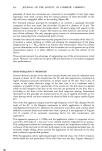
412 JOURNAL OF THE SOCIETY OF COSMETIC CHEMISTS face untreated and the other side scrubbed with an acne scrub. It is very clear that the scrubbed side is darker blue and, therefore, colder than the untreated side. The scrubbed side allows for a greater moisture loss which results in an increase in the cooling rate due to evaporation. A similar picture (Figure 11) is obtained from two arms, one untreated and the other washed five times with soap and water. Clearly the washed side is cooler. MULTISPECTRAL IMAGING OF THE SKIN I included this technique because it is a good example of how our industry can benefit from techniques used in other industries. This procedure had been used by the National Aeronautics and Space Administration for collecting information about the nature of soils, such as their water and salinity content, by using remote sensing techniques from satellites. This was further developed at the Jet Propulsion Lab of the California Institute of Technology by Dr. Anselno et al. (7) for the diagnosis of burn injuries to the skin. It has proved very valuable in categorizing the degree and depth of injury and helped in treatment decisions, such as whether or not to do a skin graft. Such decisions must be made as early as possible after the injury. On the skin, this technique involves an imaging system using computer analysis of the images. A special television camera is connected to a minicomputer. The camera obtains three images through three different filters, for example, the blue, green, and red spectral regions. Each image is stored in the computer memory. The computer is programmed to enhance the contrast of the target areas for each of the images. Then three images are treated by the computer two at a time. The ratios are computed to form three new images. The contrast of these images is again enhanced to form a false or pseudocolor final image on the T.V. monitor. The colors of this final image are used to characterize the different reflectance qualities of the target area so that areas with the same quality of reflectance have the same color. A false color image of a subject whose left side of the throat was abraded is depicted in Figure 12. This side appears to be less uniform in color than the other and lighter in appearance. This color difference is interpreted by Dr. Anselmo as indicating that the left side has experienced more irritation than the right. MACROPHOTOGRAPHY It is possible to obtain a great deal of information about the condition of the skin in vivo and about the performance of skin care products by examining photographs of the skin. As pointed out by R.V. Dent (8) in 1941, research photography is quite different from portrait photography. The latter is often concerned with the suppression of defects, while it is the purpose of scientific macrophotography to reveal them. In reviewing Dent's work, I could not help but be impressed by how complex and wonderful the organ of the skin is. The appearance of the skin to the naked eye or to the camera lens results from the way in which the different wavelengths of light are reflected from the different layers of the skin. Each layer and each component of the skin interacts with each wavelength of the spectrum of the light differently. What we see is the result of how these are allowed to penetrate, be absorbed, reflected, or
Purchased for the exclusive use of nofirst nolast (unknown)
From: SCC Media Library & Resource Center (library.scconline.org)