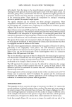
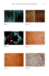
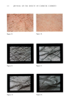
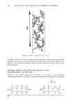
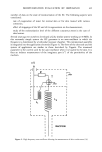
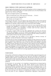
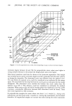
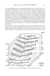
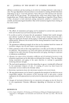
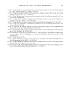
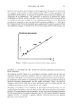
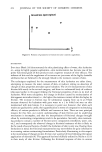
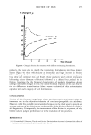
416 JOURNAL OF TIlE SOCIETY OF COSMETIC CHEMISTS Figure 19. Figure 20. Figure 21. Figure 22. Figure 23. Figure 24.

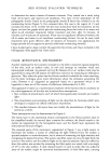
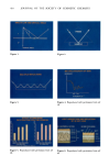
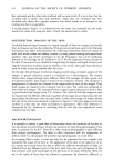
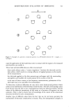
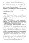
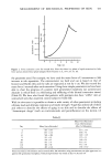
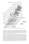
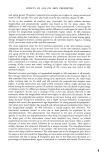
SKIN SURFACE EVALUATION TECHNIQUES 417 Figure 25. Figure 26. Figure 27. other hand, appears to have swollen. The minor divisions are more prominent with enhanced rounding in the divisional contours. CONCLUSION I would like to point out that this review of methods is far from complete. For example, one can include the Acoustic Microscopy technique where sound waves are used to reveal the structure of materials in fine detail where optical microscopes cannot see. In addition, for skin moisturization measurements, friction, transepidermal water loss and dynamic modulus are now routine tests in our laboratories. We are living in exciting times, where not only are new techniques becoming available, but some of the older ones are being upgraded to help us rapidly get more and more information about the skin.
Purchased for the exclusive use of nofirst nolast (unknown)
From: SCC Media Library & Resource Center (library.scconline.org)