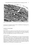
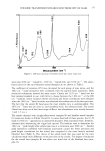
j. Soc. Cosmet. Chem., 36, 17-30 (January/February 1985) Component distributions in keratins and their estimation from amino acid analyses EMORY MENEFEE, Trichos Research, 5313 Rosalind Avenue, Richmond, CA 94805, Received October 31, 1984. Presented at the Annual Meeting of the Society of Cosmetic Chemists, New York, December 6-7, 1984. Synopsis Examination of published data supports the idea that aqueous swelling in fibrous keratins probably occurs as much in microfibrillar regions as in the matrix, except after crosslink cleavage, when the matrix becomes more hydrophilic and swellable. Approximately half the disulfide crosslinks are intramolecular, and thus not involved directly in mechanical behavior, though they may act as a reservoir for replenishing inter- molecular disulfides lost through sulfhydryl reaction. The swelling, mechanical, and reactive behavior of hair depend strongly on the location and composition of structural components, their hydrophilicity, and involvement in intermolecular crosslinking. At least four or five independent protein components are essential. Component proportions are estimated by a regression method that linearly combines the amino acids of the components to reproduce the overall amino acid analysis of the keratin. Five component proteins are taken from analyses of fractionated hair and wool: low, high, and ultra-high sulfur, cuticle, and high glycine-tyrosine. The compositions of human hair and wool samples, and certain pathological hair samples, are well-reproduced. Errors are greater when estimating compositions of other pathological hair samples or hair from lower order animals, suggesting possible requirements for the component proteins in these cases. INTRODUCTION Fifty years have passed since Goddard and Michaelis (1,2) found that two major fractions could be extracted from reduced wool fibers--one containing a higher cystine content than the other. Doubt remains, however, about how these and other components are distributed within a keratin fiber, how they contribute to the physical and chemical behavior of the fiber, or even how cystine crosslinks are connected and located. This uncertainty partly stems from the fact that about 100 distinct proteins can be isolated from a typical keratin, and that during this isolation procedure nearly all crosslinks must be broken. In recent years much of the keratin literature has supported the idea that mammalian keratins are all made up of roughly the same groups of proteins, combined differently in any particular keratin (3). This attractive notion would, if generally true, provide a very useful system by which to classify normal and aberrant keratins, and greatly simplify attempts to correlate composition with physical and chemical behavior. Un- fortunately, the fractionation procedures required for complete characterization of ker- atins are so tedious and time-consuming that few complete component analyses have 17

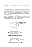
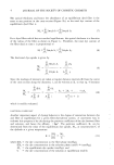
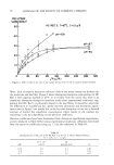
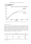
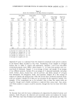
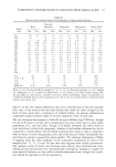
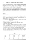
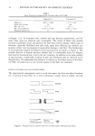
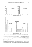
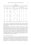
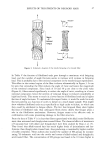
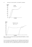
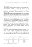
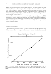
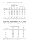
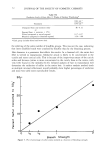
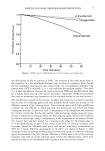
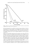
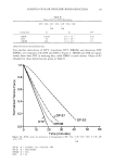
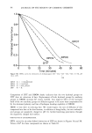
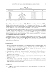
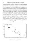
18 JOURNAL OF THE SOCIETY OF COSMETIC CHEMISTS been attempted. The present paper discusses some of the problems that still prevent full clarification of the role of crosslinks in determining keratin structure, and reports on progress of a method for mathematically isolating various components from the amino acid analyses of whole keratin fibers. THE LOCATION OF CROSSLINKS AND SWELLING The two-phase structure for keratins introduced by Feughelman (4,5) solidified for some time the assumption that nearly all aqueous swelling occurs in low-modulus matrix proteins and very little in the high-modulus microfibrils. An alternative model by Menefee (6,7) proposed a reversal of the modulus roles of the matrix and microfibrils, and also supported the idea of an ordered globular matrix, later substantiated by Spei (8,9) however, the assumption of matrix swelling was retained. Studies by Heidemann and Halboth (10), extended by Spei and Zahn (11), have strongly suggested that most swelling takes place in the microfibrils (presumably on hydrophilic side chains). Since crosslinks are so intimately involved in the valuable physical behavior of fibrous kera- tins, the extent of their involvement in a swelling phase is a crucial question.. Unfor- tunately, in spite of efforts of many years, the proper definition of a fully adequate model is still elusive. Experiments by Spei and Zahn (11,12) on the dry and wet intermicrofibrillar distances in three keratins illustrate the ambiguity of the keratin model. As seen in Table I, there is an inverse correlation between the amount of matrix and the increase in wet swelling in going from low to high sulfur keratins. Spei and Zahn interpret this by postulating that the swelling occurs chiefly in the micro fibrils. This is a reasonable idea, since the average residue side chain in the low-sulfur fraction is much more hydrophilic than in the matrix with its high proportion of cystine. On the other hand, Fraser et al. (13) claim that their method for estimating microfibril diameters in similar swollen keratins shows a microfibril wet swelling for porcupine quill of only 11%, with a matrix swelling of 53%. To account for the data of Spei and Zahn, Fraser et al. (16) present a complex and probably unnecessary mechanism. At least a portion of the differences in microfibrillar swelling spacing with different keratins is due to the effect of variations in matrix crosslink density. We can estimate the cystine content for mohair and human hair resulting from increasing the proportion of matrix from that in porcupine quills. On one extreme, if we assume that all the cystine is in the matrix (calculated to be 22.4% half-cystine for the quill), and that the increase in the total area is proportional to the squared ratios of the dry spacings, we can estimate what the cystine levels should be in mohair and human hair. As seen in the last column of Table I, they are too low even for this favorable calculation, indicating need for either a higher crosslink density in these fibers or for another higher cystine component. Choice between these alternatives is more uncertain if there is significant variation in the proportion of intramolecular-versus-intermolecular crosslinking. In a previous study (14) based on a theory of the effect of crosslinking on partial solubilization by hydrolysis, a wool sample was estimated to have 40% of its crosslinks in intramolecular form. Although other keratins were not investigated, variation in the amount of intramolec- ular-versus-intermolecular crosslinking would not be surprising. Additional indirect
Purchased for the exclusive use of nofirst nolast (unknown)
From: SCC Media Library & Resource Center (library.scconline.org)