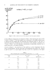
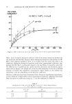
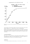
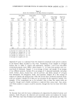
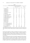
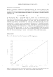
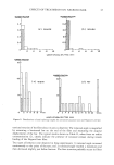
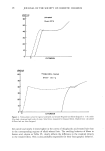
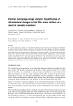
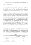
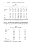
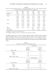
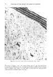
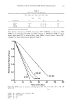
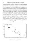
COMPONENT DISTRIBUTIONS IN KERATINS FROM AMINO ACIDS 19 Table I Changes in X-ray Spacing of Swollen Keratins (11, 12) Intermicrofibrillar 1/2 Cystine + Spacing, nm Cysteine, % Volume % Sample Matrix Dry Wet Obs Calc Porcupine quills 37 7.8 9.3 8.3 (8.3) Mohair fibers 42 8.2 9.6 10.3 9.6 Human hair 54 9.2 9.8 15.3 12.3 evidence (15,16) indicates considerable intramolecular crosslinking in fibrous keratins. The proportion of intermolecular corsslinks will have the greatest effect if swelling occurs in the matrix, since microfibrils have few disulfide crosslinks of either kind. Careful examination of spacing changes of fibers with different inter/intramolecular crosslink ratios, or of partially reduced and stabilized fibers, may offer an experimental approach toward clarifying the location of aqueous swelling. Taking into account the above ambiguities, we may attempt a sketch of a plausible model for crosslinking in fibrous keratins. Aqueous swelling may occur about as much in the crystalline slightly-crosslinked microfibril side chains as in the amorphous highly- crosslinked matrix. After cleavage of crosslinks, the matrix becomes more hydrophilic owing to the presence of sulfhydryl or cysteic acid groups. Within the matrix, crosslinks are about equally divided between intramolecular and intermolecular. Although cross- links between adjacent cystine residues are unlikely (17), the next favored configuration (16, 18) is of the form-Cys-X-Pro-Y-Cys-, with an intramolecular crosslink con- necting the two cystines. An earlier interpretation (19) of the amino acid analyses of the insoluble gel fraction remaining after partial hydrolysis of wool fibers (20) suggests that threonine, serine, valine, arginine, and glutamic acid will most often occur in the X and Y positions. This is borne out by sequence studies by Swart (21). Although intramolecular crosslinks do not directly contribute to mechanical properties, they may act as a reservoir for renewing intermolecular linkages. This is particularly important when considering how a reduced fiber may be set into a new configuration after reox- idation of sulfhydryl groups. THE MAJOR KERATIN COMPONENTS METHOD How many independent component groups are needed to define the composition and behavior of a keratin fiber? As mentioned previously, a fibrous keratin contains upwards of 100 distinct proteins, each belonging, at least partly, to some conformation family-- alpha helical chains, randomly amorphous regions, or globular structures. These pro- teins are held together by disulfide crosslinks, hydrogen bonds, and other lesser links. Attempts to build a model from this myriad detail are likely to be unsuccessful. Association of the high-sulfur fraction with the matrix, and the low-sulfur fraction with microfibrils, is currently well established. The high-sulfur (H) fraction exhibits the greatest diversity of protein content. The low-sulfur (L) fraction is much less complex. The cuticle (C) also contains a range of proteins, but on the whole resembles

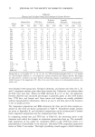
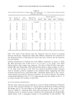
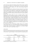
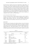
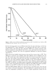
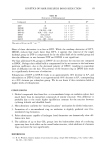
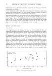
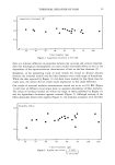
20 JOURNAL OF THE SOCIETY OF COSMETIC CHEMISTS the high-sulfur fraction. In addition, a high glycine and tyrosine (G) fraction can be isolated (22). This is thought to come from the cell membrane complex in some keratins, but occurs in greater abundance than possible from this source in others. Besides these four rough groups, an ultra-high sulfur (X) fraction has been identified (23), separable by precipitation from the high-sulfur group (24). Most fractionation studies have been carried out on whole fibers (mostly wools), so that fractions obtained represent contributions from the cortex, cuticle, and cell membrane material combined. Furthermore, there has always been some question about whether the solubilization procedure itself produces artifactive components due to chain breakage. Nevertheless, experimental studies have produced a fairly consistent set of analyses for these five components. The cuticle is included in this group because it is easily separable physically, unlike the other components, which are chemically sepa- rated. Although this may give rise to some composition overlap between the cuticle and fractionated components, the advantage of working with cuticle as though it were a component overrides this objection, at least at this stage. Table II gives mole percent analyses for each of these five components, from both human hair and wool in the L, H, and C groups, but from wool only in the X and G groups. The G group is only slightly present in hair, and the X group has so far been inadequately isolated. The hair and wool components are remarkably similar within each component group. Among the groups themselves, the X, H, and C fractions are broadly similar to each other, whereas the L and G fractions are each quite distinct. In tabulating compositions, the standard amino acid abbreviations are used, with the exception of Hcy, which is defined as the sum of half-cystine, S-carboxymethylcysteine, and cysteic acid. In what follows, the assumption is made that the amino acid composition of any human hair or wool can be approximated by a linear combination of the five components just described. To test the validity of this assumption, we examine a series of hair samples, both normal and pathological, several wools, and hair from other placental mammals, marsupials, and monotremes. Although amino acid analyses are fairly commonly done for keratins in some laboratories, the amount of reliable published data is not large. The present work should therefore be regarded as a sketch for further more careful study. The best combination of components for each keratin is found by a regression method based on minimizing the sum of squared deviations. Although this is a standard method, Gold et •l, (25) were apparently the first to propose and use it for keratin analyses, though they restricted its application to finding the best component propor- tions within a given single fractionated sample. The approach of this paper is, on the other hand, an attempt to estimate component proportions for several different keratins from a single set of components. Appendix I gives details of the method, which can be generalized to any number of components less than the number of amino acids reported in the analysis. A computer program was written in BASIC to examine all possible combinations of two or more components for goodness of fit for example, LH, LX, HC, . . . LHGC, etc. Goodness of fit, R, was defined as the RMS average of the individual deviations. Two sets of components were used: the L, H, and C analyses coming from hair samples for one set, and from wool samples for the other. Occasionally, it was found that there was some crossover, i.e., the wool set fitting hair data slightly better than the hair set.
Purchased for the exclusive use of nofirst nolast (unknown)
From: SCC Media Library & Resource Center (library.scconline.org)