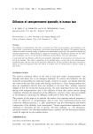
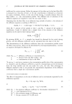
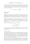
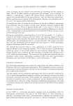
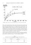
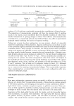
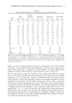
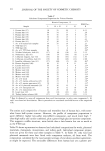
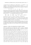
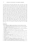
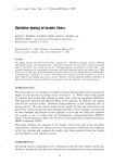
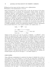
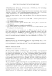
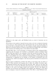
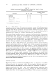
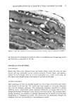
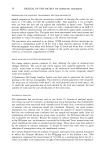
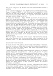
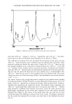
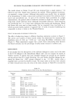
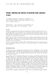
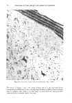
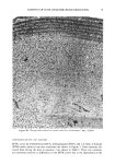
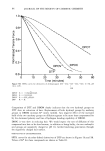
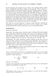
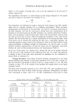
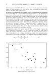
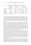
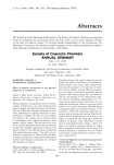
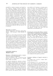
54 JOURNAL OF THE SOCIETY OF COSMETIC CHEMISTS CELL MEMBRANE COMPLEX ENDOCUTICLE • ß NUCLEAR REMNANT ß • ß MELANIN GRANULE INTERMACROFIBRILLAR MATRIX Figure 1. Diagram of non-keratinous regions of hair fiber in transverse section. from proteolytic enzymes (4), this complex is disrupted by organic solvents such as formic acid (5). Disruption of these membranes ultimately results in a complete break- down of fiber structure. Another major non-keratinous component is the endocuticle which is located at the inner portion of each cuticle layer. The endocuticle represents the remnants of the once viable cuticle cell. In the cortex, analogous components are the nuclear remnants and intermacrofibrillar matrix. These components, unlike the cell membrane complex, are composed primarily of protein and are readily digested by proteolytic enzymes. Using electron micrographs, Swift and Bews (6) showed that the liberation of cuticle cell-like units by proteolytic enzymes is due to digestion along the endocuticle sheet rather than splitting of the cell membrane complex. Similarly, they suggested that cortical cells might be liberated by digestion along the intermacrofibrillar matrix. These studies suggest that the non-keratinous components play a significant role in acting as a cement to hold the keratin fiber together. More recently, the vulnerability of these non-keratinous components to cosmetic treat- ments has been investigated. Using electron microscopy, Mahrle eta/. (7) showed the loss of substances from the endocuticle after treatment with cold-waving solutions. Similarly, Kaplin eta/. (8) reported that repetitive shampooing and drying treatments resulted in the dissolution of material from the endocuticle of hair fibers. These ob- servations were based on a subjective assessment of holes or voids appearing in electron micrographs of hair fiber cross sections. Electron microscope studies in our laboratory indicate that endocuticular voids as well as other forms of ultrastructural disruption are present in cross sections of hair fibers subjected to normal grooming processes (Fig- ure 2). We have further investigated the effects of cosmetic treatments on the ultrastructure of hair using electron microscopy in conjunction with image analysis. Our first objective was to develop a technique which would quantitatively assess ultrastructural changes

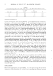
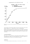
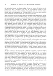
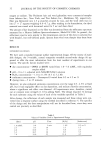
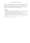
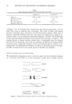
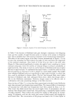
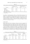
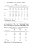
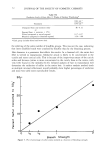
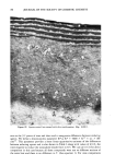
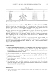
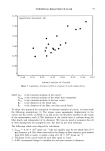
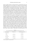
QUANTIFICATION OF ULTRASTRUCTURAL CHANGES IN HAIR 55 . ., . ,...• .: : . , .... Figure 2. Hair fiber subjected to normal grooming processes showing voids in endocuticle (50,000 x ). and then use this technique to study the effects of weathering and shampooing on the non-keratinous components of the hair. MATERIALS AND METHODS HAIR SAMPLING Brown hair fibers were obtained from one female subject whose hair had not been treated with any chemically reactive cosmetic products. Twenty fibers, each approxi- mately 18 cm in length, were cut as close as possible (1 to 2 mm) to the scalp. All hair fibers were sampled from a mid-region in the back of the head. SHAMPOO TREATMENT After sampling, the hair was divided equally into two tresses of ten fibers each which were secured at the root end. The tresses were then clipped to a one-gram laboratory hair tress for shampoo treatment. The shampooing treatment consisted of a double lathering with a conventional anionic shampoo followed by air drying. One tress was shampooed through 90 shampoo cycles during a one-week period. This multiple sham- pooing treatment was designed to simulate approximately six months of on-the-head shampooing (3.5 shampoos per week). The second tress was shampooed through one shampoo cycle and was retained as a non-shampooed control.
Purchased for the exclusive use of nofirst nolast (unknown)
From: SCC Media Library & Resource Center (library.scconline.org)