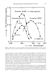
PHOTOACOUSTIC MEASUREMENTS ON SKIN 377 signal of the reference cell therefore yields the power spectrum of the light source. After narrow-band amplification, the photoacoustic signal of the skin is referenced to that of carbon black. For in vivo experiments the bottom of an open-ended cell is formed by the skin of the test subject's forearm. Self-sealing of the gas space is achieved by gently pressing the skin to the cell rim. In order to prevent overloading of the signal amplifier from bodily noise, e.g., from muscle spasms and pulsative bloodflow through subcutaneous vessels, we use a differential cell as first described by Poulet and Chambron (8). The cell has two chambers of 0.64 cm 2 cross-section area, each of which has its acoustic outlet connected with one sound port of a differential microphone (Knowles, CF 2949). A photoacoustic signal is generated only in one chamber by irradiation of the skin surface. Bodily noise is generated in both chambers. It is suppressed by the differential microphone, how- ever, as the noise signals of both chambers are almost the same with respect to magni- tude and phase. Only the signal caused by the photoacoustic effect is detected. Data acquisition and control of wavelength and chopping frequency by a microcomputer allow short measuring times. Thus burning of the skin by the high light intensities (0.3 to 3 mW/cm 2 in the wavelength range 250 to 400 nm, spectral bandwidth of 16 nm), necessary to achieve a sufficient signal-to-noise ratio, is prevented. From the stored data the differences between the photoacoustic spectra of treated and untreated skin are easily obtained. The sunscreening agents used in our study were (a) Eusolex 232 (2-Phenyl-benzimidazole-5-sulfonic acid), (b) Eusolex 6300 (3-(4-Methyl-benzylidene)-camphor) and (c) Eusolex 8020 (1-(4-Isopropyl-phenyl)-3-Phenyl-propane- 1,3-dione) from Hermal-Chemie Kurt Herrmann, 2057 Reinbek, FRG. Agents (a) and (b) are UVB filters with maximum absorption at a wavelength of 300 nm, and agent (c) is a UVA filter with maximum absorption at 340 nm. Different preparations were topically applied to the skin of the inner aspect of the forearm: Ilrido © cream, a commercially available cream of sun protection factor 3 with 2% Eusolex 232, and solutions containing 0.1 to 1% of Eusolex 6300 and 8020 in isopropanol. For the photoacoustic measurements, a volume of about 10 ptl of a given preparation was uniformly applied over a skin area of 5 X 6 cm, forming a screening layer of 3 ptm mean thickness on the surface of the skin which at the concentrations used contains amounts of sunscreening agents of 0.3 to 6 ptg/cm 2. Only a part of the incident light is absorbed within such a layer as the optical absorption lengths of the preparations, defined by the reciprocal values of optical absorption coefficients, are larger or, with the higher sunscreen concentrations and at the respective wavelengths of maximum absorption, approximately equal to the layer thickness. The topography of human skin is an obvious source of irregularities in the thickness of the applied layer. The preparations tend to accumulate in the sulci of the skin surface and form a film of reduced thickness on the plateaus between the sulci. The resulting reduction of mean opacity of the layer with the corresponding decrease in sunscreen efficacy has recently been discussed by O'Neill (9). With the topically applied isopro- panolic solutions, photoacoustic measurements were carried out after evaporation of the solvent.

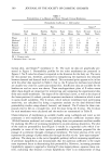
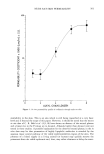
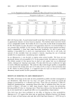
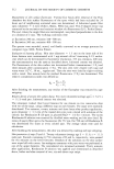
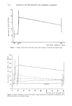
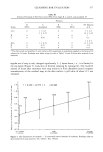
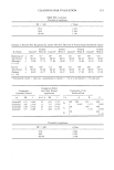
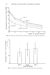
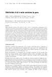
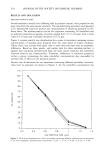
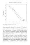
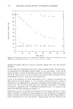
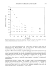
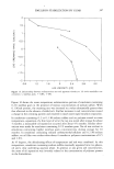
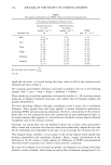
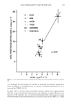
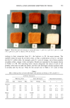
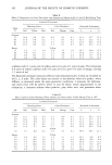
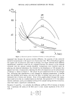
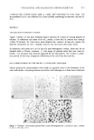
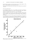
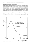
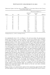
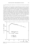
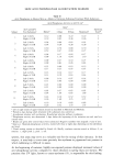
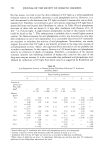
378 JOURNAL OF THE SOCIETY OF COSMETIC CHEMISTS EXPERIMENTAL RESULTS AND DISCUSSION Figure 2 shows the photoacoustic spectrum of the horny layer of a test subject's forearm in the ultraviolet spectral range, as taken in vivo. At a wavelength of 280 nm, where the aromatic amino acids of the skin proteins show maximum absorption, the optical ab- sorption length is about 4 •xm. Spectra from the skin of different test subjects are in qualitative agreement even if the signal strengths show large interindividual scatter. In a recent photoacoustic study performed on stratum corneum samples which were ob- tained by skin surface biopsy, this interindividual scatter has been shown to be caused by differences in optical and thermal properties of the superficial corneal layers (10). Table I collects in vivo data of a group of thirteen test subjects ranging in age from 24 to 28 years, measured at a chopping frequency of 1,200 Hz and a wavelength of 300 nm before and immediately after treating the inner aspect of the forearm with Ilrido © cream. At this chopping frequency the photoacoustic signal is determined with a rela- 1.0 0.5 0 I 250 300 I I PA-Spectrum of Stratum Corneum in-Vivo f = 1200 Hz I 350 nm /,00 Wavelength Figure 2. Photoacoustic in vivo spectrum of the stratum corneum of the inner aspect of the forearm. The signal amplitude is given in arbitrary units (a.u.).
Purchased for the exclusive use of nofirst nolast (unknown)
From: SCC Media Library & Resource Center (library.scconline.org)