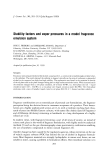
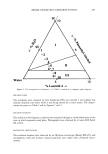
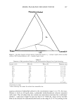
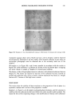
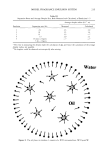
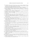
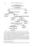
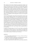
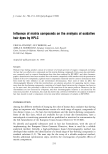
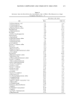
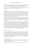
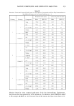
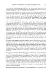
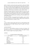
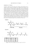
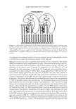
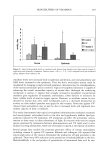
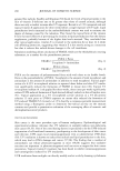
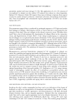
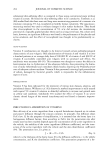
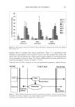
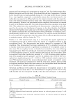
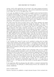
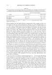
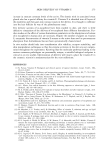
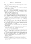
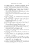
254 JOURNAL OF COSMETIC SCIENCE LO0' • Vitamin E•.X//• GS' •GSSG NADPH • '•' Vitarnin C' •+H + LOOH Vitamin E'[• •Vitamin C, GSH NADP + F{gure 5. Regeneration ooe vitarn{n E oerom (x-chromanoxy] radical Dy g]urarh]one and vitamin C. _Adapted from reference 1. glutathione peroxidase, catalase, and superoxide dismutase, as well as with smaller molecules with antioxidant properties such as ascorbic acid, glutathione, and uric acid. Direct depletion of o•-tocopherol and formation of its radical would thus affect other endogenous antioxidant pools as well. MOLECULAR MODEL OF MEMBRANE STABILIZATION BY VITAMIN E Diplock and Lucy (20) have used molecular model building to hypothesize that vitamin E may stabilize membrane structure by virtue of a specific physicochemical interaction between its phytyl side-chain and the fatty acyl chains ofpolyunsaturated phospholipids, particularly those derived from arachidonic acid. Significant interactions of o•-tocopherol with unsaturated fatty acids occur less frequently when the acyl chains of the latter are curved than when they are straight. These authors have also proposed interactions between the methyl groups of the phytyl chain of o•-tocopherol and the cis double bond of arachidonyl residues of membrane phospholipids. Thus the methyl group at C4' of o•-tocopherol can fit into a pocket provided by the cis double bond nearest the carboxyl group. The methyl group at C8' then interacts with the third cis double bond. In this "complex," the hydroxyl group of the chromanol ring of o•-tocopherol and the polar groups of the phospholipids lie together at one end, where they would be expected to participate in polar interactions at the surface of any region of membrane having a lipid bilayer structure. The fit of the methyl groups in the pockets created by the cis double bonds permits the methylene groups in the backbones of both the phytyl and fatty acyl chains to associate closely, further promoting the stability of the complex through London-Van der Waals dispersion-attraction forces. Complex formation could have the following functional consequences: (a) an inhibition of the oxidative destruction of polyunsaturated fatty acids in cells and in cellular membranes, (b) a reduction in the permeability of biological membranes, containing relatively high levels of polyunsatu- rated fatty acids, particularly arachidonic acid, and (c) the prevention of the degradation of membrane phospholipids in vivo by membrane-bound phospholipases (21-23). Simu- lation modeling, which allows calculation of lipid peroxidation indices that describe the antioxidant protection against oxidation, is available (24). Figure 6 gives a diagrammatic representation of the proposed interaction of vitamin E and polyunsaturated phospho- lipids in a biological membrane. Urano et al. (25) using •9F NMR and fluorescence polarization techniques have summarized the molecular orientation of vitamin E in liposomal membranes. They found that the chromanol moiety of o•-tocopherol was tightly adapted to the space close to the surface of membranes formed by the unsaturated fatty acid molecules in phophatidyl choline, the phytyl chain had significant motional freedom for the maintenance of a physiological fluidity, and the hydroxyl group of

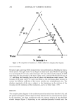
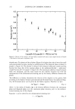
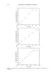
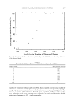
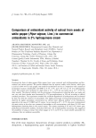
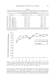
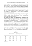
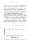
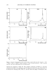
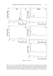
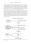
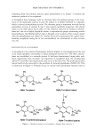
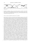
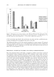
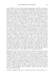
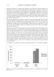
SKIN DELIVERY OF VITAMIN E 255 Antiox. idant s Figure 6. A diagrammatic representation of the proposed interactions between vitamin E, synthetic anti- oxidants, selenide- and sulphide-containing proteins, and polyunsaturated phospholipids in a biological membrane. The suggested site of action of the synthetic antioxidants is shown by the arrows. For simplicity, the phospholipids are shown as rigid structures in a bilayer membrane, but this does not imply that the membrane has a "crystalline" rather than a partially fluid character. Taken from reference 20 (with per- mission). o•-tocopherol was hydrogen bonded to the ester carbonyl group of phosphatidyl choline in membranes to retain the chromanol moiety within the space. Biological activities of some tocopherols and tocotrienols were compared to the natural (RRR) o•-tocopherol by the rat-gestation test assay. The natural o•-tocopherol was bio- logically more potent than the synthetic (a//-rac) o•-tocopherol mixture. The RRR con- figuration of the phytyl chain is optimal for biopotency (26-29). Weber eta/. (30) measured the skin penetration of a mixture of tocopherols and tocotrienols from a tocotrienol-rich palm oil fraction (TRF). After topical application of TRF, all the vita- min E forms readily penetrated into the skin of hairless mice and were present in concentrations far exceeding the baseline levels. The percent distribution of each of the vitamin E homologues in the TRF mixture was compared with its percent distribution in the skin. The percent distribution was calculated for each form as the concentration in TRF-treated skin minus the concentration in polyethylene glycol (control)-treated skin divided by the sum of the skin vitamin E concentrations x 100. They found a large fraction of o•-tocopherol absorbed into the skin compared with other forms of vitamin E. Figure 7 shows the percent distribution of vitamin E in the PEG-treated control skin, the TRF fraction itself, and TRF-treated skin after correcting for background vitamin E concentrations. The percent distribution of vitamin E forms that penetrated the skin above the background concentrations were significantly different from their distribution in the TRF mixture. Higher percentages of o•-tocopherol were found in TRF-treated skin than were present in TRF (P 0.0001), the percent of'y-tocopherol was similar to the TRF mixture, and both o•- and 'y-tocotrienols represented a smaller proportion than they did in the TRF mixture (P 0.001). The authors have suggested that the isoprenoid tail
Purchased for the exclusive use of nofirst nolast (unknown)
From: SCC Media Library & Resource Center (library.scconline.org)