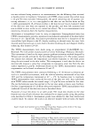
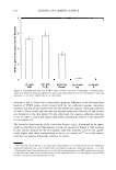
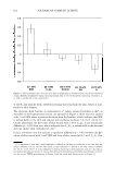
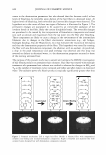
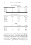
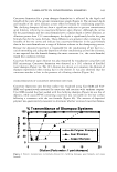
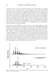
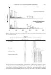
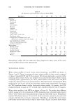
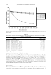
616 JOURNAL OF COSMETIC SCIENCE ous surfactant-humectant systems based on their ability to perturb the skin aqueous pores in vitro should be useful to a formulator of skin care products who is interested in understanding skin barrier perturbation induced by aqueous surfactant-humectant sys tems in vivo. The in vitro ranking methodology presented here represents a novel ap proach to rank surfactants and humectants used in skin care formulations, and may potentially reduce, or eliminate altogether, the need to conduct costly and time consuming in vivo skin barrier measurements. COMPARISON OF THE IN VITRO AND IN VIVO EFFECTS OF ADDING 10 WT% GLYCEROL TO AN AQUEOUS SDS CONTACTING SOLUTION We have recently presented an in vitro analysis that makes use of skin electrical current and mannitol skin permeability measurements to determine the effect of adding 10 wt% glycerol to an aqueous SDS contacting solution (6). Specifically, skin electrical current and mannitol skin permeability values were measured following exposure of p-FTS to an aqueous contacting solution containing SDS (1 wt%) + G (10 wt%). These values are reported again in Table III for completeness (see the in vitro results). In terms of the ranking metric (RM) analysis presented here (see the Theoretical section), a RM value of 4.1 ± 1.0 corresponds to an aqueous contacting solution containing SDS (1 wt%) + G (10 wt%), a value that is obtained by taking the ratio of the skin electrical current values in Table III corresponding to the aqueous contacting solution containing SDS (1 wt%) + G (10 wt%) (49 ± 10 µA) relative to the in vitro PBS control (12 ± 3 µA). On the other hand, Table III also shows that the RM value corresponding to an aqueous contacting Table III In Vitro and In Vivo Skin Barrier Measurements for Aqueous Contacting Solutions Containing SDS (1 wt%) and SDS (1 wt%) +- glycerol (10 wt%), Including a Comparison with the Appropriate In Vitro/In Vivo Controls Aqueous contacting solutions SDS SDS (1 wt%) + Control Skin barrier measurements (1 wt%) glycerol (10 wt%) (in vitro/in vivo)* In vitro Skin electrical current (�1A) 91 ± 10 49 ± 10 12 ± 3 Mannitol skin permeability (cm/hr) X 10 5 66 ± 10 30 ± 10 7 ± 3 Average pore radius, r 1JOI , (A) 33 ± 5 20 ± 5 20 ± 3 Enhancement in p = pjfc,** 2.6 ± 1 2.9 ± 1 1 In vivo TEWL (barrier damage) 5.30 ± 0.20 4.00 ± 0.40 3.40 ± 0.30 Visual skin dryness 0.89±0.11 0.60 ± 0.13 0.15 ± 0.02 Chromameter (erythema) 0.57 ± 0.11 0.58 ± 0.14 -0.08 ± 0.05*** *Thein vitro control corresponds to PBS in water (aqueous contacting solution iii), and the in vivo control corresponds to a no-reaction, non-occluded control (see the Experimental section). ** The enhancement in the aqueous pore number density, p, is reported relative to the in vitro control. In addition, recall that E denotes enhancer (that is, aqueous contacting solutions i, ii, iv, and v) and C denotes the in vitro control (that is, aqueous contacting solution iii). *** Note that the in vivo control shows erythema values close to zero, which is not unexpected (the small negative mean value of -0.08 results from the fact that some of the volunteers in the control group exhibited lower skin redness on Day 2, when compared to Day 1). The table reports average values and standard errors.

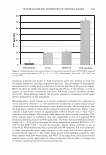
RANKING OF SURFACTANT-HUMECTANT SYSTEMS 617 solution containing SOS (1 wt%) is (91 ± 10)/(12 ± 3) = 7.6 ± 1.0, which is signifi cantly higher than the RM value of 4.1 ± 1.0 corresponding to an aqueous contacting solution containing SOS (1 wt%) + G (10 wt%). Therefore, as we have shown recently (6), adding 10 wt% G to an aqueous contacting solution containing 1 wt% SOS significantly reduces in vitro skin barrier perturbation. The mannitol skin permeability values reported in Table III also indicate a decrease in skin barrier perturbation induced by an aqueous contacting solution containing 1 wt% SOS + 10 wt% G (P = 30 ± 10 x 10- 5 cm/hr), relative to an aqueous contacting solution containing 1 wt% SOS (P = 66 ± 10 x 10- 5 cm/hr). A separate study was conducted as described earlier, comparing the effects of SOS (1 wt%) to SOS (1 wt%) + G ( 10 wt%) on the skin of healthy human volunteers to validate the in vitro model. The in vivo TEWL results in Table III indicate that the deviation from baseline in the TEWL value of skin treated with SOS (1 wt%) + G (10 wt%) is statistically lower (p 0.05) than that of skin treated with SOS (1 wt%). 14 In addition, the deviation from baseline in in vivo visual skin dryness scores in Table III reveals that SOS (1 wt%) + G (10 wt%) is not different from that of untreated skin (the in vivo control (iii)), and is statistically lower (p 0.05) in visual skin dryness than in skin treated with SOS (1 wt%). Taken together, the TEWL and visual skin dryness results reported in Table III indicate that adding 10 wt% G to an aqueous SOS (1 wt%) solution mitigates the ability of SOS to perturb the skin barrier in vivo. Using dynamic light-scattering measurements to determine the effective SOS micelle hydrodynamic radii in the presence and in the absence of 10 wt% G, we showed recently that SOS micelles are too large to penetrate into the skin aqueous pores in vitro when 10 wt% G is added to the aqueous contacting solution containing SOS (1 wt%) (6). As a result, SOS in micellar form is not able to contribute to SOS skin penetration and associated SOS-induced skin barrier perturbation. Therefore, the in vivo TEWL and visual skin dryness results reported in Table III support the in vitro results reported recently (6) by clearly showing a smaller extent of skin barrier perturbation in vivo induced by 1 wt% SOS in the presence of 10 wt% G relative to that in the absence of 10 wt% G. Although the TEWL and visual skin dryness values reported in Table III indicate that glycerol can indeed mitigate SOS-induced skin barrier perturbation in vivo, a similar corroboration was not obtained using the chromameter measurements of skin erythema (redness). This is because the chromameter scores in Table III indicate no statistical difference (p 0.05) between the erythema scores corresponding to an aqueous contact ing solution containing 1 wt% SOS in the absence of G (0.57 ± 0.11), relative to that in the presence of 10 wt% G (0.58 ± 0.14). It is possible that an aqueous contacting solution containing 1 wt% SOS + 10 wt% G may induce an initial cutaneous reaction that attracts increased blood flow to the dermis of the affected skin site, thereby leading to skin redness, even though the skin barrier may not be significantly perturbed (13- 16,19,22,29,37). Therefore, the skin at the corresponding site may appear red from an increased blood flow to the dermis, without the skin barrier being compromised, which 14 It is important to note that the deviation from baseline of TEWL corresponding to skin treated with an aqueous solution of SDS (1 wt%) + G (10 wt%) is statistically not different (p 0.05) from that corre sponding to untreated skin (the in vivo control (iii)), while it is statistically lower (p 0.05) than that corresponding to SDS (1 wt%) treated skin. This finding provides additional evidence for the ability of glycerol to mitigate SDS-induced skin barrier perturbation in vivo.
Purchased for the exclusive use of nofirst nolast (unknown)
From: SCC Media Library & Resource Center (library.scconline.org)