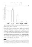
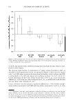
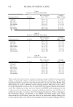
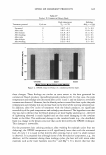
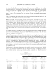
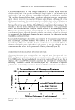
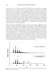
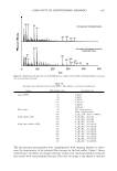
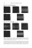
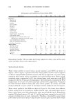
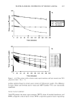
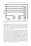
618 JOURNAL OF COSMETIC SCIENCE would lead to a higher erythema score. Accordingly, the in vivo erythema response associated with adding 10 wt% glycerol to an aqueous SDS contacting solution on the skin barrier needs to be investigated further. For example, in vivo cutaneous biochemical reaction pathways triggering erythema, which may be triggered by SDS even in the presence of glycerol, could be investigated (13,14,19,37-40). It is also possible that (a) a higher concentration of SDS than the 1 wt% used in the in vivo soap chamber, (b) a higher concentration of glycerol than the 10 wt% used in the in vivo soap chamber, and/or (c) longer exposure times of the in vivo soap chamber may be necessary to discriminate between the in vivo erythema induced by SDS in the presence of glycerol and that induced by SDS in the absence of glycerol. CONCLUSIONS Macroscopic in vitro skin barrier measurements, which quantify the extent of skin barrier perturbation induced by aqueous surfactant-humectant contacting solutions commonly encountered in skin care formulations, can be effectively used to rank these contacting solutions. Such a ranking is based on the ability of the solutions contacting the skin to perturb the skin aqueous pores of the stratum corneum (SC). An in vitro ranking metric was developed using the enhancement in the skin electrical current induced by an aqueous surfactant-humectant contacting solution relative to an in vitro PBS control aqueous contacting solution, as the metric. In vitro mannitol skin permeability measurements, when combined with skin electrical resistivity measure ments, in the context of a hindered-transport aqueous porous pathway model, provided mechanistic insight on the results of the in vitro ranking metric analysis. Specifically, the pore number density (p) and the average pore radius (r p ore ) of the skin aqueous pores induced by aqueous solutions of surfactants and humectants contacting p-FTS were determined. The in vitro skin electrical current/resistivity and mannitol skin permeabil ity measurements were carried out using the following aqueous solutions: (i) an ionic surfactant solution-SDS (1 wt%) (ii) a nonionic surfactant solution-C 12 E 6 (1 wt%) (iii) an in vitro control solution-PBS (iv) a humectant solution-propylene glycol (PG) (10 wt%) and (v) a humectant solution-Glycerol (G) (10 wt%). Utilizing the in vitro ranking metric introduced here, we obtained the following ranking order, from the mildest to the harshest, for the surfactants and the humectants considered above, based on their ability to perturb the skin aqueous pores: (v) 10 wt% G (iv) 10 wt% PG (iii) PBS (ii) 1 wt% C 12 E6 (i) 1 wt% SDS. To substantiate the findings above, in vivo soap chamber measurements were carried out on human volunteers. Specifically, the following in vivo skin barrier measurements were conducted: (a) transepidermal water loss (TEWL) determined by using an evaporimeter, (b) visual skin dryness determined by an expert grader, and (c) skin erythema measure ments using a chromameter. The overall implications of the in vivo results are that the aqueous surfactant solutions i and ii induce a larger extent of skin dryness and erythema relative to the aqueous humectant solutions iv and v. In addition, the in vivo measure ments (a-c) above indicate that a 1 wt% aqueous SDS contacting solution induces the largest extent of skin barrier perturbation, while a 10 wt% aqueous glycerol contacting solution induces the smallest extent of skin barrier perturbation. Both of these in vivo findings are consistent with the results of the in vitro ranking metric analysis. Indeed, the

RANKING OF SURFACTANT-HUMECTANT SYSTEMS 619 in vitro ranking metric predicts that adding glycerol to a solution of 1 % SDS should mitigate skin barrier perturbation. This conclusion, based on a recent in vitro study, is confirmed by the in vivo skin barrier study presented here. Therefore, determining the in vitro perturbation to the skin aqueous pores induced by aqueous surfactant-humectant systems represents a viable practical strategy to predict their in vivo skin barrier per turbation potential. The correlation established here between the in vitro ranking metric analysis, which quantifies the perturbation to the skin aqueous pores, and the in vivo skin barrier measurements can potentially be used to screen and rank many surfactants and humec tants for use in skin care formulations, thus eliminating the need to conduct costly and time-consuming testing for irritation potential. Such a practical strategy could signifi cantly speed up the effort and time required to bring new skin care formulations to the market. REFERENCES (1) P. M. Elias, and K. R. Feingold, "Skin as an Organ of Protection," in Fitzpatrick's Dermatology in General Medicine (McGraw-Hill, New York, 1999). (2) R. Scheuplein, and I. Blank, Permeability of the skin, Physiol. Rev., 51, 702-747 (1971). (3) P. M. Elias, Lipids and the epidermal permeability barrier, Arch. Dermatol. Res., 270, 95-117 (1981). (4) G. K. Menon and P. M. Elias, Morphologic basis for a pore-pathway in mammalian stratum corneum, Skin Pharmacol., 10, 235-246 (1997). (5) H. Tang, S. Mitragotri, D. Blankschtein, and R. Langer, Theoretical description of transdermal transport of hydrophilic permeants: Application to low-frequency sonophoresis, J. Pharm. Sci., 90, 545-568 (2001). (6) S. Ghosh and D. Blankschtein, The role of sodium dodecyl sulfate (SDS) micelles in inducing skin barrier perturbation in the presence of glycerol,]. Cosmet. Sci., 58, 109-133 (2007). (7) K. D. Peck, A. H. Ghanem, and W. I. Higuchi, Hindered diffusion of polar molecules through and effective pore radii estimates of intact and ethanol treated human epidermal membrane, Pharmaceut. Res., 11, 1306-1314 (1994). (8) K. D. Peck, A.H. Ghanem, and W. I. Higuchi, The effect of temperature upon the permeation of polar and ionic solutes through human epidermal membrane,]. Pharm. Sci., 84, 975-982 (1995). (9) A. Tezel, A. Sens, and S. Mitragotri, Description of transdermal transport of hydrophilic solutes during low-frequency s�nophoresis based on a modified porous pathway model,]. Pharm. Sci., 92, 381-393 (2003). (10) W. M. Deen, Hindered transport of large molecules in liquid-filled pores, AIChE ]., 33, 1409-1425 (1987). (11) J. L. Anderson and J. A. Quinn, Restricted transport in small pores, a model for steric exclusion and hindered particle motion, Biophys. J., 14, 130-150 (1974). (12) F. A. Simian, L. D. Rhein, G. L. Grove, J. M. Wojtkowski, R.H. Cagan, and D. D. Scala, Sequential order of skin responses to surfactants during a soap chamber test, Contact Dermatitis, 25, 242-249 (1991). (13) T. Agner and J. Serup, Sodium lauryl sulphate for irritant patch testing-A dose-response study using bioengineering methods for determination of skin irritation,]. Invest. Dermatol., 95, 543-547 (1990). (14) K.-P. Wilhelm, C. Surber, and H. I. Maibach, Quantification of sodium lauryl sulfate irritant der matitis in man: Comparison of four techniques: skin color reflectance, transepidermal water loss, laser Doppler flow measurement and visual scores, Arch. Dermatol. Res., 281, 293-295 (1989). (15) K.-P. Wilhelm, M. Samblebe, and C.-P. Siegers, Quantitative in vitro assessment of N-alkyl sulphate induced cytotoxicity in human keratinocytes (HaCaT)-Comparison with in vivo human irritation tests, Br. J. Dermatol., 130, 18-23 (1994). (16) K.-P. Wilhelm, A. B. Cua, H. H. Wolff, and H. I. Maibach, Surfactant-induced stratum corneum hydration in vivo: Prediction of the irritation potential of anionic surfactants,]. Invest. Dermatol., 101, 310-315 (1993).
Purchased for the exclusive use of nofirst nolast (unknown)
From: SCC Media Library & Resource Center (library.scconline.org)