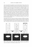
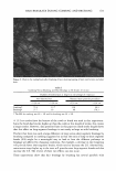
600 JOURNAL OF COSMETIC SCIENCE ment (1). Compared to the porous structure of the viable epidermis and the porous and hydrated structure of the dermis, which lie beneath the SC, the rigid and ordered structure of the SC is primarily responsible for the skin barrier function (2,3). The SC possesses an ordered brick-and-mortar structure, which consists of the flat corneocytes (the cellular bricks), interlocked with the lipid lamellae (the intercellular mortar) (1-3). The lipid lamellae of the SC consist of lipid bilayers alternating with aqueous, hydro philic layers (1-4). The observation that hydrophilic solutes are able to permeate across the SC, even under passive skin permeation conditions, has led researchers to propose the existence of tortuous, aqueous pores through the intercellular lipid lamellae in the SC. In fact, Menon and Elias (4) have established a morphological basis for the existence of aqueous pores in the mammalian SC. By applying hydrophilic and hydrophobic tracers in vivo to murine skin, under passive skin permeation conditions and under enhanced skin permeation conditions including chemical enhancers, sonophoresis, and iontopho resis, Menon and Elias visualized the resulting penetration pathways using ruthenium tetroxide staining and microwave post fixation methods (4). These aqueous pores in the stratum corneum (SC) are located in the lacunae and other aqueous regions, surrounded by polar lipids in the lipoidal mortar between the corneocyte bricks that comprise the brick-and-mortar structure of the SC (4-9). In fact, diffusion through aqueous pores in the SC has been used to explain skin penetration and the subsequent permeation of hydrophilic permeants, including mannitol (5-9), current-carrying ions like Na + and Cl - (5 ,9), and micelles of surfactants like sodium dodecyl sulfate (SDS) ( 6,29). Macroscopic in vitro skin barrier measurements, including average skin electrical resis tivity (R), which is a measure of the permeability of the current-carrying ions, and mannitol transdermal permeability (P), have been used in the context of a hindered transport theory to calculate the average pore radius (rpore) and the porosity-to-tortuosity ratio (e!T) 1 of the skin aqueous pores of the SC (5-9). Specifically, it is possible to quantify the extent of in vitro skin barrier perturbation that an aqueous solution con sisting of surfactants2 and humectants3 induces by examining the modifications of 'pore and e/T relative to an in vitro control such as PBS (phosphate-buffered saline) (5-9). The harsh surfactant SDS was shown to induce significant skin barrier perturbation in vitro relative to the PBS control, while the humectant glycerol was shown to preserve the skin barrier in vitro and to mitigate skin barrier perturbation ( 6, 12-16,21-24,30-3 5 ). Classical bioengineering techniques have been used extensively (12-16,21-24,30-35) to assess and rank the impact of surfactant solutions on human skin. However, these methods provide limited understanding of the mechanism of skin barrier perturbation. 1 The porosity, s, of the aqueous pores is defined as the fraction of cross-sectional SC area occupied by the aqueous pores, and the tortuosity, 'T, is defined as the ratio of the tortuous length of the aqueous pore to the thickness of the SC (5-9,10,11). 2 Surfactants are surface-active agents that are commonly used in skin cleansing formulations because of their ability to stabilize oil-water emulsions and clean the surface of the skin. However, they may penetrate into the skin and induce skin barrier perturbation (6,12,13,16,21,22,29). They have a hydrophilic head and a hydrophobic tail, and self-assemble to form micelles at a concentration above the critical micelle concen tration (CMC). 3 Humectants maintain the natural water content of the skin and preserve the skin barrier (30-3 5 ). In addition, some humectants like glycerol have been shown to mitigate surfactant-induced skin barrier perturbation in vitro, by preventing micelles of surfactants like SDS from penetrating into the skin through aqueous pores (6).

RANKING OF SURFACTANT-HUMECTANT SYSTEMS 601 Indeed, it would be of value to be able to correlate observations of the extent of skin barrier perturbation observed in vivo and the modification of the skin aqueous pores that an aqueous surfactant-humectant system induces in vitro relative to a control. This would allow one not only to rank these systems based on their ability to perturb the skin aqueous pores in vitro, but also to understand the mechanisms of perturbation. With the above in mind, we have developed an in vitro ranking metric that combines the characteristics of the skin aqueous pores and can potentially reduce, or eliminate alto gether, costly and time-consuming in vivo procedures such as testing for irritation potential and trial-and-error screening. Moreover, such a ranking metric would allow one to simultaneously screen and rank many surfactants and humectants for use in skin care formulations, thereby significantly speeding up the effort and time required to bring new skin care formulations to the market. In addition, a suitable ranking metric developed by measuring skin electrical currents induced in vitro relative to an in vitro PBS control can be combined with corresponding mannitol skin permeability measure ments, in the context of a hindered-transport aqueous porous pathway model. Specifi cally, use of the hindered-transport model enables the quantification of the modifications of: (i) the average pore radius, and (ii) the pore number density, induced by aqueous surfactant-humectant systems relative to the in vitro PBS control, thereby shedding light on the mechanism of in vitro skin barrier perturbation induced by the aqueous surfac tant-humectant system evaluated (see the Theoretical section). For this purpose, the anionic surfactant SDS and the nonionic surfactant C12E6 [dodecyl hexa (ethylene ox ide)}, and the humectants propylene glycol (PG) and glycerol (G), were selected (see the Materials section). The differences in the average pore radii and pore number densities induced by these two surfactants and two humectants relative to the in vitro PBS control were analyzed to gain mechanistic insight into their skin barrier perturbation/mitigation characteristics. Knowledge of the mechanism of in vitro skin barrier perturbation in duced by aqueous surfactant-humectant systems can be practically valuable in designing skin care formulations containing these chemicals that are mild to the skin and that minimize, or eliminate altogether, skin barrier perturbation (22-24,28,29,33,3 7). SDS is known to induce skin erythema (13-16,22), while C 12 E6 is known to induce skin dryness when applied to human skin in vivo (21,23,24). On the other hand, PG and G are both humectants, which are known to preserve the skin barrier and to maintain the water content of the skin when applied to human skin in vivo (30-36). The ranking of the two surfactants and the two humectants relative to the in vitro PBS control was compared with various in vivo skin barrier measurements, and the correlation between the in vitro ranking metric analysis and the in vivo skin barrier measurements was investigated. To assess the condition of the skin barrier in vivo, a soap chamber test, using a well accepted and previously published protocol (17 ,18,25-28), was utilized to treat the skin of the volar forearms of healthy human volunteers with the various surfactant-humectant solutions. Subsequently, the condition of the skin barrier was assessed by (a) transepi dermal water loss (TEWL) measurements to determine the moisture vapor flux over the skin as measured by an evaporimeter, which serves as an excellent quantitative indicator of skin barrier perturbation in vivo (25-27), (b) visual skin dryness scores determined by an expert grader to clinically assess the extent of skin dryness (17 ,23 ), and (c) skin erythema (redness), objectively determined by chromameter measurements (18). The
Purchased for the exclusive use of nofirst nolast (unknown)
From: SCC Media Library & Resource Center (library.scconline.org)