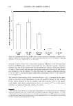
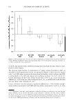
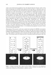
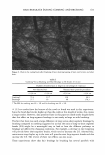
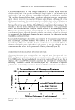
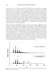
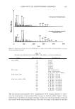
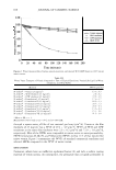
602 JOURNAL OF COSMETIC SCIENCE results of these in vivo measurements are reported here, and have been compared with the in vitro measurements. To further validate the in vitro ranking metric, we compared the in vitro and in vivo skin barrier mitigation effects of adding glycerol to SDS aqueous contacting solutions (6). This comparison demonstrates the ability of glycerol to mitigate skin barrier perturba tion upon exposure to SDS solutions, which provides evidence of the ability of glycerol to prevent SDS micelles from penetrating into the SC and inducing skin barrier per turbation in vivo. This important finding is fully consistent with our recent finding on the ability of glycerol to hinder SDS micelle penetration into the skin in vitro by reducing the average radius and the number density of the skin aqueous pores (6). EXPERIMENT AL IN VITRO SKIN BARRIER STUDIES Materials. Sodium dodecyl sulfate (SDS) was purchased from Sigma Chemicals (St. Louis, MO). Analytical-grade glycerol (G) and propylene glycol (PG) were purchased from VWR Chemicals (Cambridge, MA). 3 H-radiolabeled mannitol was purchased from American Radiolabeled Chemicals (St. Louis, MO). Dodecyl hexa (ethylene oxide), C 12 E6, was purchased from Nikko Chemicals (Tokyo, Japan). All these chemicals were used as received. Water was filtered using a Millipore Academic water filter (Bedford, MA). Phosphate-buffered saline (PBS) was prepared using PBS tablets from Sigma Chemicals (St. Louis, MO) and Millipore filtered water, such that a phosphate concen tration of 0.01 M along with a NaCl concentration of 0.137 M was obtained at a pH of 7.2. Preparation of the solutions. For the in vitro skin barrier studies, the following aqueous solutions were prepared: (i) an anionic surfactant solution-SDS (1 wt%) (ii) a nonionic surfactant solution-C 12 E6 (1 wt%) (iii) a control solution-phosphate-buffered saline (PBS) (iv) a humectant solution-propylene glycol (PG) (10 wt%) and (v) a humectant solution-glycerol (G) (10 wt%). Preparation of the skin samples. Female Yorkshire pigs (40-45 kg) were purchased from local farms, and the skin (back) was harvested within one hour after sacrificing the animal. The subcutaneous fat was trimmed off using a razor blade, and the full-thickness pig skin was cut into small pieces (2 cm x 2 cm) and stored in a -80°C freezer for up to two months. The in vitro experiments involve contacting pig full-thickness skin (p-FTS) with aqueous surfactant-humectant and PBS control solutions i-v, and were performed according to previously published protocol (5 ,6,29). Mannitol trans dermal permeability measurements. Vertical Franz diffusion cells (Permegear Inc., Riegelsville, PA) were used for the mannitol skin permeability measurements (5,6,29). All the experiments were performed at room temperature (25°C). The p-FTS samples were mounted in the diffusion cells with the SC facing the donor compartments. Both the donor and the receiver compartments were filled with PBS, and the p-FTS samples were left to hydrate for one hour before the beginning of the experiment to allow the skin's initial barrier property to reach steady state (5,6,29). At this point, the skin electrical current across the p-FTS sample was measured, and only p-FTS samples with an initial skin current 3 pA were utilized in the mannitol skin permeability studies

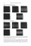
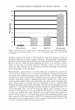
RANKING OF SURFACTANT-HUMECTANT SYSTEMS 603 (5,6,29). PBS in the donor compartments was then replaced separately with 1.5 ml of aqueous contacting solutions i-v (5 ,6,29), and left in contact with the SC of the p-FTS samples for five hours (6,29). Subsequently, aqueous contacting solutions i-v were removed and the donor compartments along with the p-FTS samples were rinsed four times with 2 ml of PBS to remove any trace chemical left on the skin surface and in the donor compartments. Subsequently, the p-FTS samples in the diffusion cells were ex posed to aqueous contacting solutions of 3 H-radiolabeled mannitol in PBS (1-10 µCi/ ml) for 24 hours (5 ,6). For additional experimental details, including a discussion of the liquid scintillation counting method used to determine the mannitol transdermal per meability, see references 5, 6, and 41. Skin electrical current and resistivity measurements. During each mannitol transdermal (skin) permeability experiment, two Ag/AgCl electrodes (E242, In Vivo Metrics, Healdsburg, CA) were placed in the donor and receiver compartments to measure the electrical current and the electrical resistivity across the p-FTS samples (5,6). A 100-m V AC voltage (RMS) at 10 Hz was generated by a signal generator (Hewlett-Packard, Atlanta, GA) and was applied across the two electrodes for 5 s. The electrical current across the skin was measured using an ammeter (Hewlett-Packard, Atlanta, GA). The electrical resistance of the p-FTS sample was then calculated from Ohm's law (5,6,41). The skin electrical resistivity was obtained by multiplying the actual skin electrical resistance by the skin area (A = 1.77 cm2) (5,6,41). Skin electrical current and resistivity measure ments were carried out before and during the permeation experiments at each prede termined sampling point. For each p-FTS sample, an average skin electrical resistivity was determined over the same time period for which the steady-state mannitol skin permeability, P, was calculated (5,6,41). IN VIVO SKIN BARRIER STUDIES The objective of this study was to conduct quantitative in vivo skin barrier perturbation measurements upon contacting volar forearm skin test sites of human volunteers with aqueous solutions i, ii, iv, and v. The control for these in vivo measurements was an untreated skin test site that was not occluded and that was not exposed to aqueous contacting solutions i-v. This control will be referred to hereafter as the in vivo control to differentiate it from the in vitro PBS control (iii). Note that this in vivo control was adopted because studies have shown that natural hydration of the skin in vivo can be mimicked in vitro by contacting skin with a PBS solution that has a pH of 7-7.4 (the in vitro control, which is similar to the in vivo pH of the hydrated SC (13-17)). The in vivo skin barrier perturbation studies were carried out using a modified soap chamber test (12,17 ,18). The modified soap chamber test involved application of patches con taining aqueous contacting solutions i, ii, iv, and v to skin test sites on the volar forearms of 96 female volunteers (four groups of 24). The volunteers were interviewed to verify that they had no known allergies to soaps or fragrances, and that they were not using any medications that could have interfered with the results of the study. 4 Following the enrollment of the volunteers, the condition of the skin barrier of the volar forearm test 4 Note that the equivalent in vitro selection criterion used was a skin electrical current 3 µA at the beginning of the study (6,41).
Purchased for the exclusive use of nofirst nolast (unknown)
From: SCC Media Library & Resource Center (library.scconline.org)