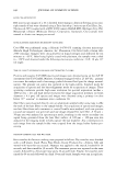
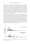
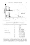
]. Cosmet. Sci., 58, 663-666 (November/December 2007) Abstracts International Journal of Cosmetic Science Vol. 29, No. 4, 2007* Contact Angle Measurement - A Reliable Supportive Method For Screening Water-Resistance Of Ultraviolet Proceting Products In Vivo R. Hagens, T. Mann, V. Schreiner, H. G. Barlag, H. Wenck, K.-P. Wittem and W. Mei Beiersdorf AG, Research Bioengineering, Unnastra8e 48, D- 20245 Hamburg, Germany Ralf Hagens, Beiersdorf AG, Research Bioengineering, Unnastra8e 48, D-20245 Hamburg, Germany Substantivity of sunscreen formulations is affected by the wash-out rate of ultraviolet-absorber and -reflector compounds in water. Water-resistance of sunscreen formulations is currently determined according to a standardized European Cosmetic Toiletry and Perfumery Association (COLIPA) protocol, encompassing the determination of a minimal erythemal dose before and after a defined immersion step in water. It can be supposed that the higher the wettability of a treated skin area, the higher is the wash-out rate of sunscreen compounds. This present report addresses the validity of determining the wettability of treated skin alone as a measure for the water-resistance of sunscreen products. The report addresses the robustness, accuracy and congruence of a recently developed wettability test, based on the measurement of the contact angle (CA) of a sessile water drop on treated skin areas. Contact angle data of 66 sunscreen formulations are compared with the corresponding results of 81 water-resistance tests, using the sun protection factor (SPF)/immersion/SPF method. Sunscreen products tested by the CA method were applied to the skin of the volar forearm of test subjects at a defined dose and drying-time, using a standardized application and recording device. Contact angles between a sessile water drop and skin were recorded by a Charge-Coupled Device (CCD) camera and subjected to automatic contour analysis. Taking the SPF/immersion/SPF method as gold standard, accuracy parameters of the CA method were determined. By using an appropriate cut-off level of CAs, the CA method has a specificity and positive-predictive value of 100%, and turns out to be a reliable screening method to identify water resistant formulations. Based on our findings, those formulations that give CAs above 30° may be categorized water-proof without further testing by the COUP A water resistance method. In Vitro human Nail Penetration and Kinetics of Panthenol X. Hf*, S. B. Homby t , R. C. Wester*, S. Barbadillo*, Y. Appa and H. Maibach* *Dermatology, University of California San Francisco, Surge 110, Box 0989, 9 f Medical Center Way, San Francisco, CA 94143 and 'Neutrogena Corp., 5760 West 96ht Street, R&D Building, Los Angeles, CA 90045, U.S.A. Dr Xiaoying Hui, Department of Dermatology, University of California San Francisco, Surge 110, Box 0989, 90 Medical Center Way, San Francisco, CA 94143, U.S.A. The in vitro absorption of panthenol into and through the human nail was examined in this study. Panthenol, the alcohol form of pantothenic acid (vitamin BS), is believed to act as a humectant and improve the flexibility and strength * These abstracts appear as they were originally published. They have not been edited by the Journal of Cosmetic science. 663

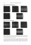
664 JOURNAL OF COSMETIC SCIENCE of nails. A liquid nail treatment formulated with panthenol efficacy and good tolerance of C8-LHA make it an (2%) was compared to a solution of panthenol (2%) in water. excellent candidate for the treatment of hyperpigmentory Fingernail specimens were dosed daily for 7 days with either disorders. the nail treatment (non-lacquer film forming) formulation or aqueous solution with sampling performed every 24 h. Panthenol concentrations were determined in the dorsal surface, interior (by drilling and removal) and in the supporting bed under the human nail. Panthenol levels in the dorsal nail (R2 = 0.87 P 0.001), nail interior (R2 = 0.94 P 0.001) and nail supporting bed (R2 = 0.79 P 0.003) showed a significant linear increase with each day of dosing. Significantly more panthenol was delivered into the interior nail and supporting bed by a nail treatment formulation than from an aqueous solution. The film acts not only as a reservoir of panthenol, but also acts to increase the hydration of the nail and the thermodynamic activity of panthenol as well, thereby enhancing diffusion A Simple Experimental Method To Study Depigmenting Agents M. L. Abella, J. de Rigal and S. Neveux L'Oreal Recherche, Chevilly-Larue, France Dr M. L. Abella, Centre de Recherche L'Oreal, 188 rue Paul Hoebart, 94550 Chevilly Larue, France. The first objective of the study was to verify that a controlled UV exposure of four areas of the forearms together with randomized product application enabled to compare treatment efficacy and then to compare the depigmenting efficacy of different products with a simple experimental method. Sixteen volunteers received 0.7 minimal erythermal dose for four consecutive days. Products tested were ellagic acid (0.5%), vitamin C (5%) and C8-LHA (2%). Product application started 72 h post last exposure, was repeated for 42 days, the control zone being exposed, non-treated. Colour measurements included Chromameter®, Chromasphere®, Spectro-colorimeter and visual assessment. Comparison of colour values at day I and at day 7 showed that all zones were comparably tanned, allowing a rigorous comparison of the treatments. We report a new simple experimental model, which enables the rapid comparison of different depigmenting products. The Review Article: Nail Biology And Nail Science D. A. R. de Berker*, J. Andret and R. Baran* *Bristol Dermatology Ce r, e, Bristol Royal Infirmary, Bristol BS2 8HW, U.K., Department of Dermat � ogy, CHU Saint Pierre, 1000 Brussels, Belgium and Nail Disease Centre, 42, rue des Serbes, 06400 Cannes, France Robert Baran, Nail Disease Centre, 42, rue des Serbes, 06400 Cannes, France The nail plate is the permanent product of the nail matrix. Its normal appearance and growth depend on the integrity of several components: the surrounding tissues or perionychium and the bony phalanx that are contributing to the nail apparatus or nail unit. The nail is inserted proximally in an invagination practically parallel to the upper surface of the skin and laterally in the lateral nail grooves. This pocket-like invagination has a roof, the proximal nail fold and a floor, the matrix from which the nail is derived. The germinal matrix forms the bulk of the nail plate. The proximal element forms the superficial third of the nail whereas the distal element provides its inferior two-thirds. The ventral surface of the proximal nail fold adheres closely to the nail for a short distance and forms a gradually desquamating tissue, the cuticle, made of the stratum comeum of both the dorsal and the ventral side of the proximal nail fold. The cuticle seals and therefore protects the ungual cul-de-sac. The nail plate is bordered by the proximal nail fold which is continuous with the similarly structured lateral nail fold on each side. The nail bed extends from the lunula to the hyponychium. It presents with parallel longitudinal rete ridges. This area, by contrast to the matrix has a firm attachment to the nail plate and nail avulsion produces a denudation of the nail bed. Colourless, but translucent, the highly vascular connective tissue containing glomus organs transmits a pink colour through the nail. Among its multiple functions, the nail provides counterpressure to the pulp that is essential to the tactile sensation involving the fingers and to the prevention of the hypertrophy of the distal wall tissue, produced after nail loss of the great toe nail.
Purchased for the exclusive use of nofirst nolast (unknown)
From: SCC Media Library & Resource Center (library.scconline.org)