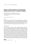
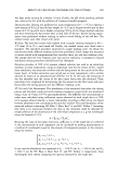
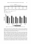
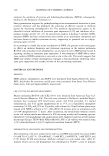
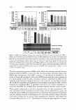
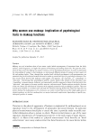
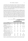
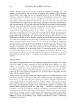
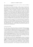
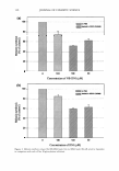
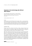
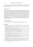
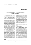
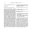
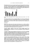
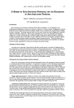
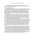
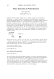
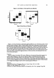
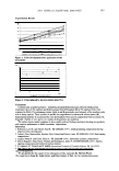
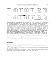
2007 ANNUAL SCIENTIFIC MEETING 18 16 E E 14 ... 12 0 .. .! 10 I C 8 6 0 In vivo experiments: Dose-dependent effect of SSR on Langerhans cells In ex vivo skin treated with white tea ■\8hicle • \\hite tea 2 3 4 SSR do• (J/cm2) Figure 1 This study aimed to compare the effect of topical formulations containing T4 Endonuclease liposomes, RNA fragments, or a lotion containing white tea on the prevention of UV-induced contact hypersensitivity suppression in humans. 169 100 subjects ofFitspatrick Skin type I to III and MED ranging between 20-50 mj/cm2 were randomized into 5 groups which received no treatment, vehicle, or one of the above preparations. Products were selfapplied on gluteal skin once daily for 6 days. Halfofthe subjects per group received solar simulated radiation at 0. 75 MED, over the treatment site on day 3 of product application. DNCB sensitization followed 3 days after irradiation. The contact hypersensitivity response was measured 2 weeks after sensitization by the increase of skin fold thickness over 5 elicitations on the arm. Effect of a 0.2% white tea preparation on the UV-induced immunosuppression: Results clearly demonstrate that the treatment with the white tea containing product resulted in a significant protection against the UV-induced langerhans cells depletion (22% reduction in CD la positive cells after UV exposure and treatment with the white tea, vs. 57% reduction after UV exposure alone), and as a result prevented most of the UV-induced Contact Hypersensitivity suppression (27% reduction in CHS after UV exposure and treatment with the white tea, vs. 53% reduction in CHS after exposure to UV light alone). Similarly, the experiments conducted with a preparation containing either 1 % micrococcus lysate (T4 endonuclease), or 1% RNA fragments, both provided protection from UV-induced Langerhans cell depletion as well as UV-induced Contact Hypersensitivity Suppression (63% reduction of CHS after exposure to UV alone vs. 18% reduction after UV and treatment with the Micrococus lysate). Conclusions: The combination of the Ex vivo and In vivo experiments clearly demonstrate the effect of protecting the skin celJs from oxidative damage as well as increasing the natural DNA repair process results in the prevention the UV-induced immuno suppression. This effect seems to originate from the inhibition of the UV-induced migration of the Langerhans cells away from the skin. Clearly, the combination of this technology with sunscreens enhances significantly the protective benefits provided by the latter. These technologies altogether will allow for the development of products which will provide a far broader protection than what is obtained actually with topical sunscreens formulations.

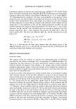
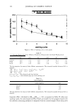
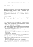
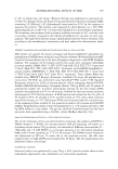
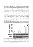
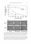
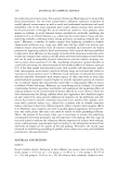
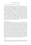
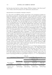
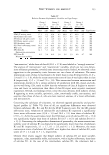
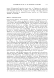
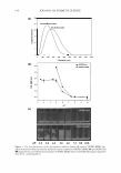
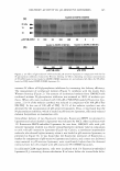
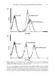
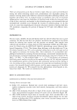
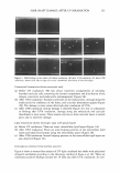
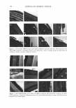
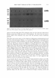
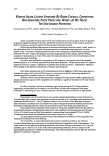
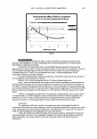
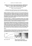
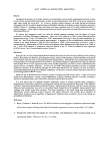
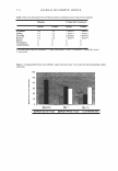
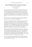
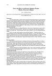
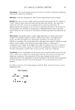
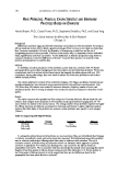
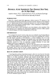
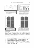
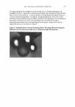
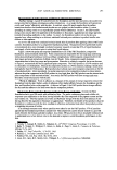
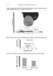
170 JOURNAL OF COSMETIC SCIENCE Do WE ALSo FACE HAZARDS TRIGGERED By Sus .. ERVTHEMAL UV EXPOSURE] RESULTS OF A CLINICAL STUDY ON VARIOUS BIOLOGICAL ENDPOINTS Jurgen Vollhardt1, Ph.D., Antony R. Young2, Guy Orchard2, Graham I. Harrison2 and Jochen Klock1 1 DSM Nutritional Products, R&D Personal Care, P.O. Box 3255, 4002 Basel, Switzerland 2 St. John's Institute of Dermatology, Lambeth Palace Road, London SE1 7EH, United Kingdom Repeated sub-erythemal UV exposure on human skin may occur during short periods of UV exposure while performing non extensive outdoor activities. This may also occur during extensive UV radiation exposure while on holidays through improper use of sun protection combined with the use of low protection factors. The effects of such repeated radiation are barely investigated. There are no visible signs of skin redness in this case, however, we were asking: is there significant damage going on which would call for intervention? In particular we were interested whether the immune status of the skin is already compromised after sub-erythemal radiation and if this could be prevented through application of sunscreens. We studied the effects of 11 consecutive daily sub-erythemal exposures of solar simulated radiation (SSR) on buttock skin of 6 healthy sun sensitive skin types 1/11 (18-35 years). A standard dose was given for each exposure which represented 0.52 or 0.65 minimal erythema doses (MED) depending on the MED of the volunteer. Erythema was assessed daily and biopsies were taken to assess end-points relevant in regards to the formation and propagation of skin cancer. We also evaluated the effects of a broad-spectrum (4* UVA) daily-care low SPF (7.5) sunscreen with 6% Polysilicone-15 (PARSOL ® SLX) as a UVB filter and 2% Butyl Methoxydibenzoyl-methane (PARSOL ® 1789®) as a UVA filter. The study, approved by the Ethics Committee of St Thomas' Hospital London, was done according to the Declaration of Helsinki. For 11 consecutive days 6 volunteers were exposed each day to a radiation of 0.6 MED. Biopsies were taken at day 0, 5, 11 and 12. CD1 a served as marker to investigate the presence of Langerhans cells (LC). In addition, we checked skin redness, quantified thymine dimers, p53 expression, the proliferation markers MIB-1, and searched for the indicators of apoptosis: BCL-2 and sun burned cells (SBC). There where two test sites on the skin: one with and one without a broad spectrum sunscreen applied. Results Erythema accumulated on the vehicle control site but not on the sunscreen site (Figure 1 ). At day 12, the vehicle site showed a dramatic increase in the erythema index and considerably more DNA damage than the sunscreen site (Figure 2). Overall, sunscreen treated sites showed much less DNA damage than the vehicle sites at all time-points (p 0.01 - 0.03). 1 2 , . 1 , 1 Day Figure 1 Erythema Index t I 10 11 12 Figure 2: lmmunostaining of thymine dimers. a: placebo control, b: sunscreen protected SC: stratum comeum, E: epidennis D: dennis n: nuclear staining
Purchased for the exclusive use of nofirst nolast (unknown)
From: SCC Media Library & Resource Center (library.scconline.org)