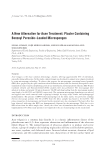
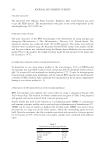
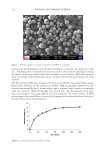
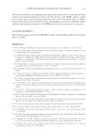
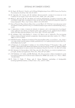
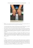
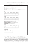
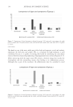
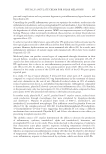
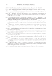
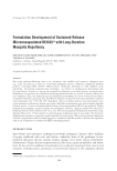
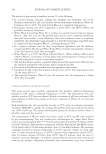
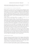
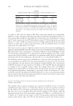
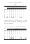
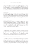
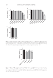
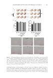
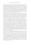
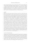
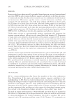
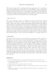
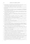
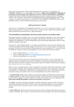
128 JOURNAL OF COSMETIC SCIENCE with IPL for better aesthetic results. The exclusion criteria included: participants who were smokers, diabetic, presented precancerous lesions during dermatoscopy, and had large discrepancy of lesions between the hands. Dermatoscopy was performed in all participants, with a Dermlite 3 Gen® portable dermatoscope with diode light and 10-times magnification, to rule out possible associations with precancerous lesions. Melanosis was marked on both the right and left hands of the 60 participants, with macules as similar as possible in color and diameter on both hands. This marking was performed to serve as a parameter for the analysis of erythema and temperature when the participants return after 48 h. This melanosis was properly identified in the photographs to facilitate findings at the time of reassessment Figures 1 and 2. Figure 1. G1 participant. (A) photo before treatment, (B) photo immediately after with the marking of the melanosis of both hands in which the measurement of perilesional erythema in mm was performed with the dermatoscope and where the temperature was also measured, and (C) 48 h after treatment where higher erythema can be seen on the left hand. Figure 2. G2 participant. (A) photo before treatment, (B) photo immediately after with the marking of the melanosis of both hands in which the measurement of the erythematous halo in mm was performed with the dermatoscope and where the temperature was also measured, and (C) – photo 48 h after treatment where a more intense inflammatory process is observed in the right hand with edema and blisters.

129 PHYSALIS ANGULATE CREAM FOR SOLAR MELANOSIS The participants were divided into two groups (for each group, n = 30). In group 1 (G1), we compared the differences in the improvement of IPL-driven inflammation in the hand where the cream with 0.5% P. angulata L. extract was applied with the contralateral hand where 1% hydrocortisone was applied. In group 2 (G2), we compared one hand treated with P. angulata extract with the other hand that was treated only with the vehicle (placebo). All participants signed the Informed Consent Form (ICF) before treatment. CREAM MANIPULATION The supercritical extract of P. angulata (already patented under the name Physavie®) was supplied by the company Chemyunion. The manipulation of the cream containing the vegetal extract followed the criteria provided by Chemyunion, and the 0.5% concentration was indicated by the manufacturer. The other creams used followed the conventional rules for manipulation. The vehicle was the same in all preparations (lanette cream – Mapric®). One hundred and twenty bottles of cream were manipulated, each weighing 40 g. G1 received 30 bottles with 40 g of 0.5% P. angulata L. extract and lanette cream identified with a red label and 30 bottles with 40 g of 1% hydrocortisone and lanette cream identified with a yellow label. G2 received 30 bottles with 40 g of 0.5% P. angulata L. extract and lanette cream identified with a green label and 30 bottles with a blue label containing 40 g of the lanette cream only (the placebo). The identification of each bottle, with the respective color of each label and the decision of which bottle would go to which group, was random and made by the responsible pharmacy. APPLICATION OF INTENSE PULSED LIGHT The participants were photographed with a 12-megapixel digital camera under the black background and then treated with IPL using a 540-nm wavelength filter that is absorbed by melanin. This is the most suitable treatment for melanosis. The Etherea® machine from the company Vydence, serial code 06154-14, Anvisa Registration 800585800-15, was used. Light was emitted by a sapphire tip (4.2 cm long and 1.5 cm wide) at a speed of one flash per second using a 540-nm wavelength filter. This wavelength is absorbed by melanin, making it the most suitable treatment. The energy parameters used were based on the phototype indicated by the manufacturer and the existing guidelines in the literature (1–22). Phototype II, III, and IV participants were treated with energy of 18–20 J/cm2 and a pulse duration of 10 ms, 17–20 J/cm2 and a pulse duration of 10 ms, and 15–17 J/cm2 and a pulse duration of 15 ms, respectively. In G1 and G2, most participants were phototype III (86.6%). IPL was applied by placing the sapphire tip directly in contact with the skin and pressing the trigger button on the handpiece where the 540-nm IPL filter was attached. Immediately after pressing the button, the beam was emitted at the speed of a flash per second, and this entire area of the skin was treated. Then, the tip was placed immediately next to where the previous shot was taken to treat the new area. This was performed successively from the thenar to the hypothenar region, and from the wrist toward the fingers, until the entire area received the emission of the light beams equally, thus treating the entire hand. The number of shots depended on the size of each hand and did not influence the results. It
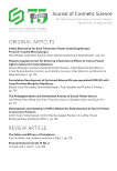
Purchased for the exclusive use of nofirst nolast (unknown)
From: SCC Media Library & Resource Center (library.scconline.org)