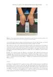
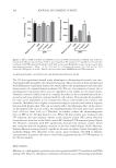
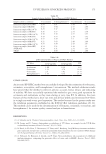
136 JOURNAL OF COSMETIC SCIENCE Presence of polyunsaturated fatty acids (omega-3, omega-6, and omega-9) was revealed previously in the same study. Polyunsaturated fatty acids have an important role in the restoration of skin barrier healing because they modulate inflammatory responses (32). Dermatological procedures such as IPL, laser, and peeling can cause damage to the skin surface, which compromises the skin barrier function as these methods remove epidermal cells (33–35). There is evidence that the skin barrier is metabolically active and participates in inflammatory responses through the activation of cytokines, fibroblasts, melanocytes, and new vascular formation (36). Omega-9 fatty acid can significantly inhibit the production of nitric oxide, improve the level of interleukin-10, and decrease the expression of cyclooxygenase-2 (COX-2) in the skin lesion (37–39). It is possible that the improvement of inflammation through P. angulate does not happen only by using physalins and phytosteroids. The present study showed similar results to those of previous studies conducted using P. angulate. G1-PA results were less effective than G1-H. In contrast, G2-PA showed better results than G2-V such as improvements in all objective and subjective parameters including pain, erythema, and temperature. The temperature in 28 participants (93.3%) was higher in the hand that was treated with the moisturizing extract of P. angulate, and in 13 participants (43.3%), it was 1°C. CONCLUSION This study shows that the cream containing 0.5% P. angulate L. extract may be useful when a mild anti-inflammatory agent is required for treating skin-related inflammations. Although the response was lower compared to hydrocortisone, these clinical results highlighted the anti-inflammatory properties of the cream and its potential as a dermocosmetic. This study also paves the way for further research involving preclinical studies with different concentrations to prove the benefits and quantify the anti-inflammatory action of P. angulata in comparison to that of corticosteroids. REFERENCES (1) S. Cignachi, V. Campos, L. Maluf, L. Grohs, M. Wancizinski and M. Costa, Comparative study of the effectiveness of 2940-nm, 1340-nm laser and intense pulsed light use on global rejuvenation of hands. J Am Acad Dermatol, 72, 1–267 (2015). (2) A. Kawada, H. Shiraishi, M. Asai, H. Kameyama, Y. Sangen, Y. Aragane and T. Tezuka, Clinical improvement of solar lentigines and ephelides with an intense pulsed light source. J Dermatol Surg, 28 (6), 504–508 (2002). (3) H. Sasaya, A. Kawada, T Wada, A. Hirao, and N. Oiso, Clinical effectiveness of intense pulsed light therapy for solar lentigines of the hands. J Dermatol Surg, 24 (6), 584–586 (2011). (4) R. C. R. Patriota, C. J. Rodrigues, and L. C. Cucé, Luz intensa pulsada no fotoenvelhecimento: Avaliação clínica, histopatológica e imuno-histoquímica. An Bras Dermatol, 86 (6), 1129–1133 (2011). (5) M. G. Cunha, F. D. Paravic and C. A. Machado, Alterações histológicas dos tipos de colágeno após diferentes modalidades de tratamento para remodelamento dérmico: Uma revisão bibliográfica. Surg Cosmet Dermatolo, 7 (4), 285–292 (2015). (6) S. A. P. Sampaio and E. A. Rivitti, Dermatologia. 3rd Ed. (Artes Medicas, São Paulo, 2008).

137 PHYSALIS ANGULATE CREAM FOR SOLAR MELANOSIS (7) V. Campos, A. Filippo and R. Matos, “Rejuvenescimento com Lasers e Outras Fontes de Luz,” in Rotinas de Diagnósticos e Tratamento da Sociedade Brasileira de Dermatologia, O. Lupi, J. Belo, and P. Cunha. Ed. (Guanabara Koogan, Rio de Janeiro, 2010) pp. 415–417. (8) P. Babilas, S. Schreml, R. M. Szeimies, and M. Landthaler, Intense pulsed light (IPL): A review. Lasers in Surgery and Medicine: The Official Journal of the American Society for Laser Medicine and Surgery, 42 (2), 93–104 (2010). (9) Y. Lin, H. C. Chiang, W. S. Kan, E. Hone, S. J. Shih and M. H. Won, Immunomodulatory activity of various fractions derived from Physalis angulata L. extract. Am J Chin Med 20 (3–4), 233–243 (1932). (10) B. J. M. Silva, S. W. G. Pereira, A. N. D. Rodrigues, J. L. M. Nascimento and E. O. Silva, In vitro antileishmanial effects of Physalis angulata root on Leishmania infantum. J Integr Med, 16 (6), 404–410 (2018). (11) C. P. Sun, C. Y. Qiu, F. Zhao, N. Kang, L. X. Chen and F. Qiu, Physalins V-IX, 16,24-cyclo-13,14-seco withanolides from Physalis angulata and their antiproliferative and anti-inflammatory activities. Sci. Rep, 7 (1), 4057 (2017). (12) T. C. B. Tomassini, N. S. Barbi, I. M. Ribeiro and D. C. D. Xavier, Gênero Physalis – uma revisão sobre vitaesteróides. Quim Nova, 23 (1), 47–57 (2000). (13) D. C. D. X. P. Lopes, Z. M. F. Freitas, E. P. Santos and T. C. B. Tomassini, Atividade antimicrobiana e fototóxica de extratos de frutos e raízes de Physalis angulata L. Rev Bras Farmacogn, 16 (2), 206–210 (2006). (14) G. N. T. Bastos, A. R. S. Santos, V. M. M. Ferreira, A. M. R. Costa, C. I. Bispo, A. J. A, Silveira and J. L. M. Nascimento. Efeito antinociceptivo do extrato aquoso obtido de raízes de Physalis angulata L. em camundongos. Journal of ethnopharmacology, 103 (2), 241–245 (2006). (15) N. B. Pinto, T. C. Morais, K. M. B. Carvalho, C. R. Silva, G. M. D. Andrade, G. A. D. C. Brito and F. A. Santos, Topical anti-inflammatory potential of Physalin E from Physalis angulata on experimental dermatitis in mice. Phytomedicine, 17(10), 740–743 (2010). (16) W. A. S. Cruz, “Atividade de mieloperoxidase e produção de oxigênio singlete em neutrófilos e células monocíticas.” Dissertation, Universidade de São Paulo – USP. 2010. (17) N. A. V. D. E. O. Sharma, A. Bano, H. S. Dhaliwal and V. Sharma, A pharmacological comprehensive review on “Rassbhary” Physalis angulata (L.). International Journal of Pharmacy and Pharmaceutical Sciences, 7(8), 34–38. (2015) (18) K. Basey, B. A. Gray and J. G. Wooley. Phygrine, and alkaloid from physalis species. Phytochemistry, 31, 4173–4176 (1992). (19) N. Ismail and M. Alan, A novel cytotoxic flavonoid glycoside from Physalis angulata L. Fitoterapia, 72, 676–679 (2001). (20) C. D. C. P. Lopes, E. P. Santos, T. C. B. Tomassini, Atividade anti-séptica de formulações contendo extrato etanólico de frutos de Physalis angulata L. Ver Bras Farmacogn, 86 (2), 75–77 (2005). (21) J. T. Checon, “Atividade anti-inflamatória do extrato liofilizado de Physalia Angulata L. em cultura de queratinócitos humanos e seu potencial como ativo dermocosmético.” Dissertation, Accra: Universidade Estadual Paulista, Botucatu – SP. 2011. (22) S. F. Hengeltraub and E. O. Monteiro, Avaliação da atividade anti-inflamatória do Umiditá AI creme e do Umiditá AI loção em cultura de queratinócitos humanos normais expostos a agentes surfactante irritante. Ver Bras Med, 71, 5–12 (2015). (23) C. Isaac, P. R. S. Ladeira, F. M. P. Rêgo, J. B. B. Aldunate, R. M. C. Tutihashi and M. C. Ferreira, Alterações no processo de reparo fisiológico. Rev Bras Queimaduras, 10 (2), 61–65 (2011). (24) J. L. Shupack, K. Washenik and G. H. Pak, “Glicocorticoides,” in Fitzpatrick Tratado de Dermatologia. 5th Ed. T. B. Fitzpatrick, I. M. Freedberg, A. Z. Eisen, et al. Ed. (McGraw Hill, Rio de Janeiro, 2005), pp. 2713–2717. (25) K. Breuer, T. Werfel and A. Kapp, Safety and efficacy of topical calcineurin inhibitors in the treatment of childhood atopic dermatitis. Am J Clin Dermatol, 6, 67–77 (2005).
Purchased for the exclusive use of nofirst nolast (unknown)
From: SCC Media Library & Resource Center (library.scconline.org)