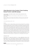
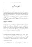
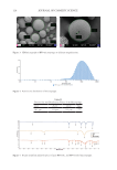
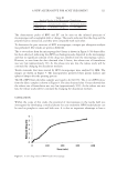
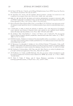
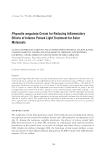
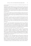
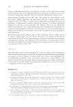
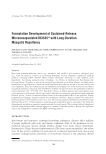
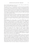
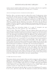
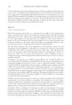
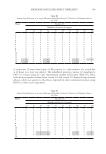
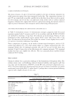
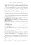
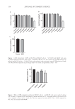
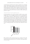
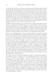
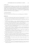
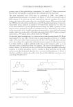
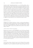
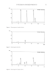
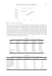
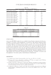
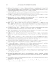
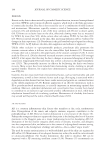
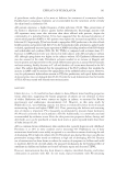
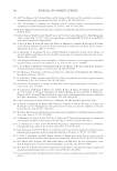
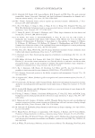
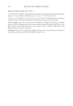
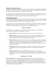
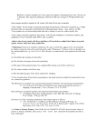
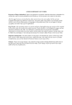
158 JOURNAL OF COSMETIC SCIENCE Contro l 0.0625 mg /m L 0.12 5 mg / mg/m L mg/m L 1 mg2 / mg/m L 4 m g/mL 0 50 100 Co n t l 0. 625 m g/mLmg/m 0. 1 2 L 0.25 m g/mL 0 mg/m L 1 m g/mLg/mL 2 m mg/m L 0 50 100 a b Control UVB 0 50 100 c Figure 1. (A,B) Cytotoxicity of SFE on B16-F0 and HaCaT cells (n = 5). B16-F0 and HaCaT cells were incubated without (control) or with 0.0625–4 mg/mL of SFE for 24 h. (C) Cytotoxicity of UVB radiation (50 mJ/cm2) on HaCaT cells (n = 3). HaCaT cells were irradiated with or without (control) UVB radiation (50 mJ/ cm2). Results were expressed as percentages relative to control and presented as mean ± SD. Negati ve controArbuti2 l n mg /m L 1 mg/m L 0 50 100 *** *** *** Figure 2. Effect of SFE on melanin synthesis in B16-F0 cells (n = 4). B16-F0 cells were incubated without (NC) or with 1–2 mg/mL of SFE, and arbutin 1 mg/mL was used as PC. Percentage values in the treated cells were compared to those in the control cells. Data are expressed as mean ± SD ***p 0.001 compared to NC. NC: cells treated with DMEM. B16 Cell viability% HaCaT Cell viability% HaCaT cell viability % Melanin content %

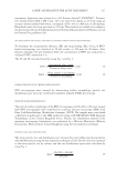
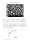
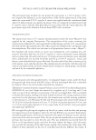
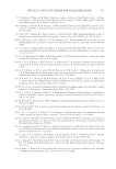
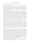
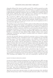
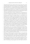
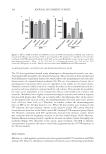
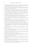
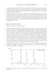
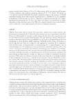
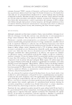
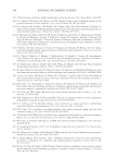
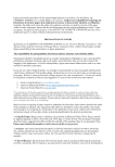
159 ANTIPIGMENTATION AND ANTIOXIDANT ACTIVITY in the hydroxylation of monophenol and the oxidation of o-diphenol to the corresponding o-quinone, which ultimately is converted to melanin through several reactions. As shown in Figure 3, SFE (1 mg/mL) significantly decreased the tyrosinase activity by 27.3%, compared with the control. The obvious impact on tyrosinase was observed after 48 hours of treatment. PC arbutin (1 mg/mL), as expected, weakened the enzyme expression, but not as much as SFE. However, the tyrosinase activity of SFE at the concentration of 2 mg/mL reduced by 20.6%, and such a phenomenon was consistent with melanin inhibition. The aforementioned results showed that the optimal concentration of SFE to inhibit melanin generation is 1 mg/mL. ANTIOXIDANT EFFECT OF SFE AGAINST UV-TREATED HACAT CELLS To examine antioxidant capacity of SFE, the T-AOC and GSH-Px activities in UVB- damaged HaCaT cells were investigated. Statistically, there was a marked difference of T-AOC activity between the UVB model group and the control group (Figure 4A, p 0:05). Different concentrations of SFE and vitamin C (0.1 mg/mL) were cultured with HaCaT cells for 6 hours and exposed to UVB radiation (50 mJ/cm2). SFE (1 mg/mL) significantly elevated the T-AOC activity to 0.060 mmol/g, which was better than PC vitamin C. Compared with the control group, GSH-Px activity in the UVB model group was obviously decreased (Figure 4B, p 0.01). Surprisingly, SFE at the concentration of 2 mg/mL remarkably increased GSH-Px activity, while it showed no effect on GSH-Px activity at the concentration of 1 mg/mL. The reason for these differences remains unclear. Overall, these results revealed that SFE could considerably protect the antioxidant enzyme activities in UVB-damaged HaCaT cells and further restrain UVB-induced oxidative stress. Negati ve controArbu l ti 2 mg/m1 L mg/m L 0 50 100 *** ** * Figure 3. Inhibitory effect of SFE on intracellular tyrosinase activity (n = 4). Different concentrations of SFE and 1 mg/mL of PC arbutin were incubated with intracellular tyrosinase and L-DOPA at 37°C. Tyrosinase activity was measured by the change in absorption at 490 nm. Results were expressed as percentages of control. Data were presented as mean ± SD *p 0.05, **p 0.01, and ***p 0.001 compared to NC. NC: cells treated with DMEM. Tyrosinase content %
Purchased for the exclusive use of nofirst nolast (unknown)
From: SCC Media Library & Resource Center (library.scconline.org)