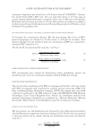
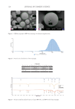
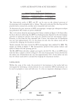
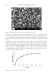
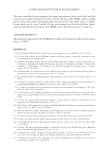
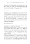
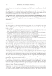
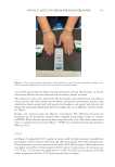
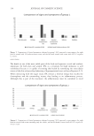
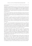
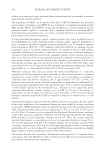
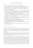
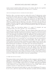
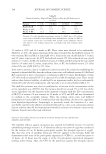
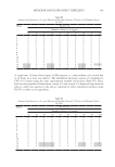
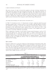
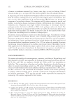
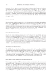
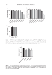
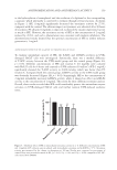
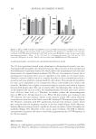
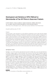
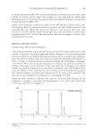
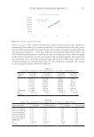
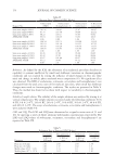
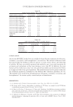
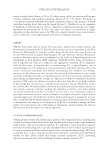
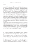
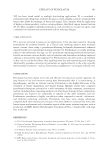
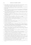
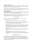
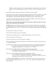
160 JOURNAL OF COSMETIC SCIENCE 3D SKIN EQUIVALENT TO INVESTIGATE THE WHITENING EFFICACY OF SFE The 3D skin equivalents showed many advantages in dermatological research over two- dimensional (2D) monolayer cell cultures for bioassay. This is because of their stratified and well-differentiated epidermal barrier (18), which reflects the morphological and molecular characteristics of a normal human epidermis (19). The use of reconstructed tissues also is advantageous in situations where an active ingredient is not soluble in cell culture media. Therefore, it may be soluble in an oil or a complex base that can be accommodated easily by a reconstructed tissue platform compared with the cell cultures. This provides the possibility for more active ingredients to be evaluated for efficacy and broadens the cosmetic raw materials. MelaKutis has a highly consistent histological structure and melanin response function with human skin. This can accurately reflect the whitening effect of the extract on the human body and can realize the multidimensional detection (gene level, protein level, cell level, tissue level, etc.). Therefore, we further evaluate the antimelanogenesis effect of SFE in the 3D skin model in vitro. When 3D skin models were irradiated with UV radiation, the total melanin content in the negative control (NC) group obviously increased more than that in the blank control (BC) (without UVB exposure) group (Figure 5C). However, treatment with SFE significantly decreased the melanin content, which was consistent with the brightness variation in the skin model (Figure 5A-B). Moreover, Fontana–Masson staining revealed a significant decrease in melain content throughout the epidermis (Figure 5D), efficiently in the cuticle, upon treatment with SFE at different concentrations. In general, the extract could reduce pigmentation due to UVB exposure in the 3D skin models. DISCUSSION Melanin, as a dark pigment, provides protection against harmful UV irradiation and DNA damage (20). However, abnormal accumulation of melanin causes dermatological problems Negati ve contro l UV B Vitamin C 2 mg/m L 1 mg/m L 0.00 0.02 0.04 0.06 # * * UVB Negati ve c o n t l UVB Vi t in C 2 mg/m L 1 mg/m L 0 2000 4000 6000 ## ** * UVB a b Figure 4. Effects of SFE on T-AOC and GSH-Px activities in UVB radiation-induced HaCaT cells. Cells were treated with different concentrations of SFE (2 and 1 mg/mL) or PC vitamin C (0.1 mg/mL) for 24 h and then irradiated with UVB radiation (50 mJ/cm2). (A) T-AOC activity and (B) GSH-Px activity and were tested. Data were shown as the mean ± SD (n = 3) ## p 0.01 and # p 0.05 versus the control **p 0.01 and *p 0.05 versus the UVB group. As an NC, HaCaT cells were treated with DMEM without UVB irradiation. T-AOC/(mmol/ GSH-Px/(U/mg protein)

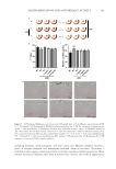
161 ANTIPIGMENTATION AND ANTIOXIDANT ACTIVITY including melasma, senile lentigines, and skin cancer (21). Melanin synthesis involves a series of enzyme-catalyzed and nonenzyme-catalyzed chemical reactions. Tyrosinase, a multifunctional copper-containing enzyme, positively regulates melanin generation. Many natural tyrosinase inhibitors have been discovered for cosmetic and medical applications. Figure 5. UVB-exposed MelaKutis was treated with 500 µg/mL kojic acid or different concentrations of SFE (2, 1, 0.5 mg/mL). (A) Photograph of MelaKutis was presented on day 7. (B) The intensities of pigmentation on day 7 were measured by a colorimeter, and data were expressed as the L* values. (C) Melanin content on day 7 was tested. (D) 3D skin models cultured for 7 days were subjected to Fontana–Masson staining. Data were represented mean ± SD from three independent samples ## p 0.01 and # p 0.05 versus the BC group **p 0.01 and *p 0.05 versus the NC group. (BC: without UVB exposure, NC: UVB exposure, PC: UVB exposure + kojic acid.) White arrows: melanin.
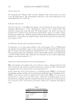
Purchased for the exclusive use of nofirst nolast (unknown)
From: SCC Media Library & Resource Center (library.scconline.org)