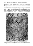
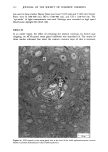
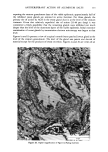
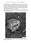
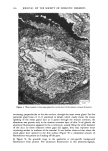
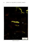
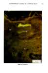
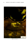
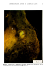
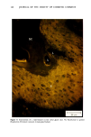
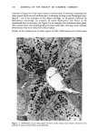
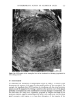
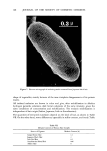
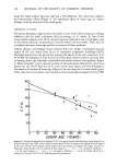
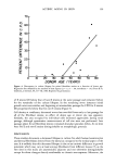
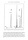
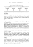
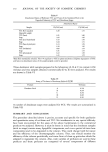
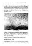
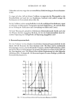
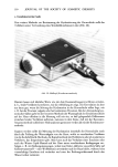
ANTIPERSPIRANT ACTION OF ALUMINUM SALTS 131 glands were also performed. Figure 10 is a cross-sectional view of a typical secretary coil of an untreated gland, demonstrating the characteristic clear and dark cells, secretary granules, etc. At the time of biopsy (about 15 min after the subjects left the environmental chamber), these glands were probably still secreting. An example of an ACH-treated sweat gland is portrayed in Figure 11. The lumens of treated glands, interestingly, were nearly completely devoid of the products of sweat secretion. No evidence of gross cellular disturbances, membrane damage, etc. was observed. An example of the intradermal duct of an untreated sweat gland is presented in Figure 12. At this level, the products of sweat secretion in the lumen were still readily .... • Figure 12. Intradermal duct region of a water-treated eccrine sweat gland (L:lumen).

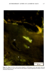
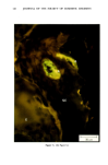
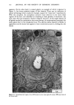
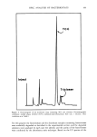
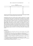
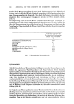
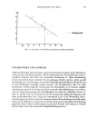
132 JOURNAL OF THE SOCIETY OF COSMETIC CHEMISTS apparent. On the other hand, in treated glands, an example of which is depicted in Figure 13, the lumen remained empty of that material. There was no indication of ductal wall damage which would, for example, support the "leaky hose" hypothesis (6). With one exception, the amorphous occlusive mass observed in the more distal regions of the duct, and identified as consisting at least partially of aluminum, was never seen. The one exception is shown in Figures 14a and b. In this single instance of all glands studied by transmission electromicroscopy, the antiperspirant penetrated the sweat gland duct to the intradermal level. The electron dense material is fibrillar in appearance and the ductal wall appears to have constricted around it, swelling shut the lumen. Figure 15. Intradermal duct region of an ACH-treated eccrine sweat gland (L:lumen DW:outer limit of the ductal wall).
Purchased for the exclusive use of nofirst nolast (unknown)
From: SCC Media Library & Resource Center (library.scconline.org)