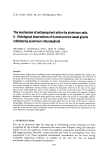
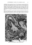
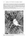
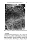
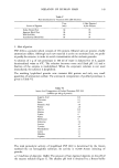
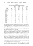
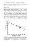
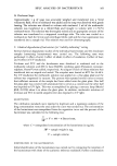
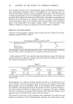
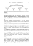
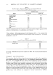
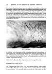
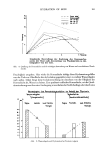
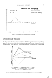
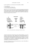
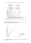
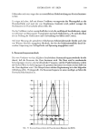
ACTINIC AGING IN SKIN 159 • 60 5o 4o 30 I% 20 ß ß ß ß ß ß ß t 20 30 40 50 60 70 80 DONOR ,ZiGE ('YEARS) Figure 4. Discrepancy in culture lifespan for paired fibroblast strains as a function of donor age. Regression line obtained by the method of least squares (y = 1.2 x - .48, correlation = .75). (From B. A. Gilchrest,J. Gerontol., 35, 537-541, 1980. Reprinted by permission.) of E strains fell below that of non-E strains at the same passage and remained below for the remainder of the culture lifespan. In the remaining seven instances initial growth rates were similar, and beginning at intermediate passage the CPD for E strains fell progressively below that for non-E strains (Figure 5). Cell density at confluency decreased more than two-fold from early to late passage for all of the fibroblast strains no effect of donor age or donor site was apparent. Similarly, the area occupied by individual cells increased appreciably during serial passage, although quantitative measurements of cell size were not performed. Late passage plates for all fibroblast strains contained abundant granular debris. At no time were the E and non-E strains distinguishable on morphologic grounds. DISCUSSION These studies document a decreased lifespan in culture for adult human keratinocytes and dermal fibroblasts derived from the lateral as compared to the medial aspect of the arm. It is unlikely that this decreased lifespan is due to an intrinsic difference in growth potential, which may vary at least among fibroblasts from different tissues (7), as the two sites in this study are anatomically adjacent and not otherwise distinguishable except for those changes directly attributable to chronic sun-exposure. Moreover, the

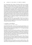
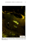
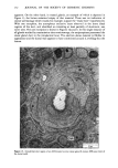
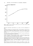
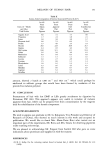
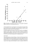
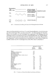
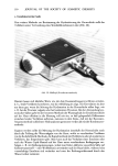
160 JOURNAL OF THE SOCIETY OF COSMETIC CHEMISTS 50 x x I' 40 - 30- 20- •.• - 71 year 01U •r I0 • ' non-E . .'.., .... 0 5 I0 15 20 40[ . ioL -./• 41 yeor old donor i i i i I i ß ß ß I , . , , I ß ß , ß I. 0 5 I0 15 20 PASSAGE LEVEL Figure 5. Plot of cumulative population doubling versus passage level for fibroblast strains derived from the medial, non-exposed (non-E) and lateral, sun-exposed (E) aspects of the upper arm of two adult donors. Cells were passaged at approximately 2 week intervals during exponential growth phase and at 3 to 4 week intervals during late passage. Paired horizontal bars indicate the 2 CPD determinations made from parallel plates at each passage. (From B. A. Gilchrest, J. Gerontol., 35,537-541, 1980. Reprinted by permission.) percent reduction in lifespan for both keratinocytes and fibroblasts derived from the lateral aspects of the arm appears to increase as a function of donor age, an effect difficult to explain with this hypothesis. Rather, it seems likely that a cumulative environmental influence is responsible for the observed difference. While it is not possible in an experiment of nature to isolate or quantitate each variable, it is probable that the different growth capacities observed in the paired keratinocyte and fibroblast strains reflect the two donor sites' different cumulative exposure to the sun, and specifically to ultraviolet light, that portion of the sun's
Purchased for the exclusive use of nofirst nolast (unknown)
From: SCC Media Library & Resource Center (library.scconline.org)