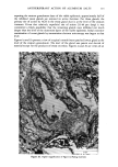
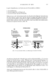
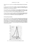
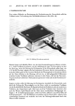
ACTINIC AGING IN SKIN 155 Fibroblast Cultures The dermal strip was cut into 1 mm 2 fragments and 2 or 3 fragments were separately and firmly applied to a scored Falcon culture dish and moistened with 1 ml of medium for 6 to 24 h, at which time an additional 3 ml of medium was gently added. The medium was subsequently changed weekly until the fibroblast outgrowth covered at least half the dish. At this point, cells were rinsed with EDTA 0.2%, incubated with trypsin .25% for 3 to 5 min and then dislodged from the dish by brief vigorous pipetting. The resulting cell suspension was centrifuged, resuspended in fresh medium, counted in a hemocytometer chamber, and replated at a density of 105 cells in a 60-ram dish with 4 ml medium. Subsequently the plates were examined twice weekly and the medium changed if nonconfluent. Subcultures Keratinocytes were subcultured at three- to four-week intervals, when individual colonies were large and the supporting 3T3 feeder layer had begun to detach spontaneously from the dish. Fibroblasts were subcultured at approximately two week intervals, as soon as confluence was attained. Standard techniques were used (2,3). Definitions Plating efficiency (keratinocytes only): the number of colonies visible on a stained plate after three weeks growth, divided by the total number of cells originally plated. Average number ofcellsper colony (CPC). After removing fibroblasts from the plate, the remaining keratinocytes are trypsinized and counted, and this number is divided by the number of colonies counted on a paired stained plate. Cumulative population doublings (CPD). In the case of keratinocytes, if one assumes that each colony arises from a single progenitor cell, the average number of cells per colony equals 2 c after G generations and hence G = ln(CPC)/ln 2. In the case of fibroblasts, G = in (N/No)/ln 2, where N = final cell count and No -- initial cell count, a formula which assumes exponential growth of all plated cells. Cumulative population doublings (CPD) = G in the case where all plated cells divide exponentially throughout the passage. CPD is the more accurate term to be used in the situation where not all cells behave in this manner and the number of generations (G) can be determined only for an average cell, not for each cell in a population. Senescence. Keratinocytes were judged to be senescent when only small abortive colonies could be seen after three to four weeks cultivation, and further subculturing was impossible. Fibroblasts were judged to be senescent when plates innoculated at standard densities, initially capable of achieving confluence in less than two weeks, were no longer confluent after four weeks. RESULTS At the clinical level, all subjects exhibited striking differences between the chronically sun-exposed and non-exposed aspects of the upper arm. Skin on the outer aspect was more deeply wrinkled, darker and "thicker," or more difficult to pinch between two

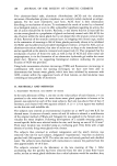
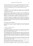
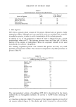
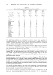
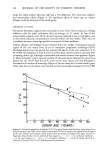
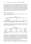
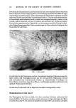
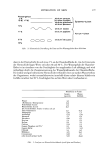
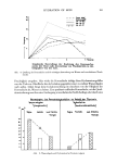
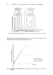
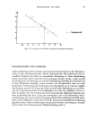
156 JOURNAL OF THE SOCIETY OF COSMETIC CHEMISTS 50- 40- 30 - 10- 20 30 40 SO 60 70 80 AGE (YEARS) Figure 1. Differences in plating efficiency for primary cultures of keratinocytes from chronically sun-exposed (E) and non-exposed (non-E) sites, E/non-E, as a function of donor age. Note 11 to 32.5-fold difference. Regression line obtained by the method of least squares (y = .Sx -- 9, correlation coefficient = .83). (From B. A. Gilchrest,J. Invest. DermatoL, 72, 219-223, 1979. Reprinted by permission.) fingers while the inner aspect was pale and smooth in all subjects. Skin obtained from the outer aspect of the arm also had more telangiectasia, irregular hyperpigmentation, and benign epidermal proliferation such as seborrheic keratoses, although no patient had lesions compatible with actinic keratoses, basal cell carcinoma, or squamous cell carcinoma. These clinical changes were most prominent in the 70 to 80-year-old subjects and least prominent in the two 28-year-old subjects. KERATINOCYTE CULTURES Keratinocyte strains from both the sun-exposed and nonexposed aspects of the arm were successfully carried to senescence in six of the twelve subjects. In the remaining cases, one or both of the cell strains was lost at early passage because of bacterial contamination. In the successful cultures, stratified epidermal colonies were first visible at the 8 to 32-cell stage, 8 to 12 d after initial plating. Most colonies grew in an orderly manner over the subsequent two to three weeks, remaining circular in configuration with tightly adherent cells. On most plates there was a wide range of colony size so that after three weeks colonies containing more than 1,000 cells were intermixed with abortive colonies of fewer than
Purchased for the exclusive use of nofirst nolast (unknown)
From: SCC Media Library & Resource Center (library.scconline.org)