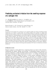
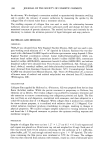
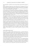
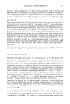
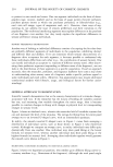
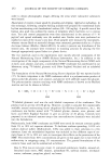
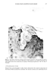
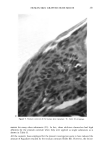
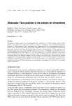
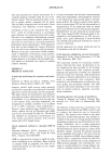
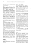
HUMAN SKIN GRAFTED NUDE MOUSE 257 Figure 4. The athymic nude mouse stratum corneum and part of epidermis. The loosening of the layers is frequently observed due to the technical difficulties of slide preparation. For thickness measurement the most compact section of the corneum layer was employed. Microscopic magnification x 400.4C. After 15 strippings. AUTORADIOGRAPHIC STUDIES Results from autoradiographic studies clearly demonstrate that selected components of the Natural Moisturizing Factor permeate the stratum corneum and underlying cells of

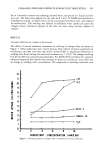
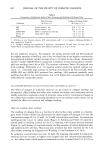
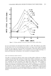
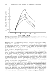
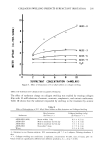
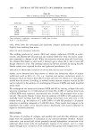
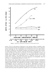
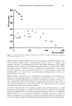
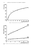
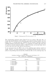
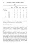
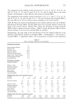
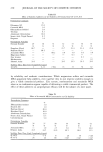
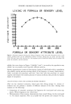
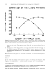
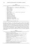
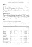
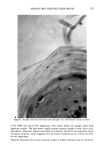
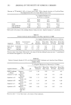
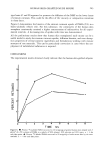
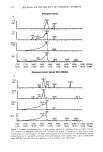
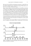
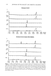
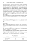
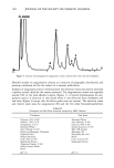
258 JOURNAL OF THE SOCIETY OF COSMETIC CHEMISTS the epidermis. This can be seen in Figure 2, which is a photomicrograph of a section of grafted human skin which was treated with a •4C-labeled amino acid, biopsied, and autoradiographed by the technique described. The juncture between the basal cells of the epidermis and the dermis is not clearly visible due to the fact that staining proce- dures, which involve treatment with aqueous solutions of dyes, were not performed due to the possibility that this would leach the water-soluble labeled components from the microtomed section. However, the stratum corneum is clearly visible and the basal layer can be approximated as the lower level of the dark-colored portion of the skin. Other components of the Natural Moisturizing Factor produced comparable results. RESULTS OF THE PRELIMINARY STRIPPING EXPERIMENTS Verification of the efficacy of stripping procedure. The rate of removal of the stratum corneum of the human skin transplant is shown in Figure 3. The results obtained correlate closely with the results reported by Schaeffer et alo (19). It is easily observed from the figure that the loosely attached upper layers of the stratum corneum can be removed easily by employing consistent stripping pressure. The amount removed decreases at the lower layers. This may affect the amount of recovery of the applied material in each layer. The optimization of the stripping technique was determined by examining the stripped stratum corneum under the microscope (Figures 4 and 5). In Figure 4A, the non-stripped mouse stratum corneum is shown. The stripping theoretically should re- move the horny cells layer by layer. However, this removal was not continuous but patchwise as was reported earlier (19). Stratum corneum after being stripped five times is shown in Figure 4B. Although the stratum corneum layers were loosened during the preparation for microscopic examination, uneven removal of the corneum is noticeable. In Figure 4C a section of mouse stratum corneum, which was stripped 15 times, shows deep surface ridging and the hairshaft openings are filled heavily with stratum corneum (20). This can create an uneven thickness of the horny layer. Therefore, twenty strip- pings were found necessary to remove all the remaining horny cells. At this level some of the stratum granulosum was damaged by this procedure. Figure 5A illustrates the full thickness stratum corneum of the human skin transplant. The stripping procedure reduces the amount left on the epidermis, as shown in Figure 5B. However, one or two layers of corneocytes may remain in patches after twenty strippings, as shown in Fig- ure 5C. Microscopic examination of the mouse skin and transplants revealed that the mouse stratum corneum consisted of about 7 to 12 layers and human corneum of about 12 to 23 layers. Since the average number of layers in abdominal skin ranges between 16-20 (6), a possible thickening of the corneum of the transplant may occur. Tape stripping analyses. Results of tests to determine the efficiency of the tape-stripping method demonstrated that essentially all of the applied dose of •4C-labeled substances could be recovered by the cleansing and tape-stripping procedures, provided these pro- cedures were carried out within a few minutes after application to the skin (Table I). Results of stripping of sites dosed with test substances clearly demonstrate that selected components of the Natural Moisturizing Factor permeate into and through the stratum corneum as shown in Tables II and III. Table II illustrates the stratum corneum uptake of some of the single major components
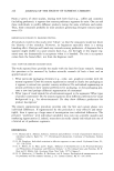
Purchased for the exclusive use of nofirst nolast (unknown)
From: SCC Media Library & Resource Center (library.scconline.org)