j. Soc. Cosmet. Chem., 39, 347-354 (November/December 1988) The role of human melanin in providing photoprotection from solar mid-ultraviolet radiation (280-320 nnl) N. KOLLIAS and A. H. BAQER, Physics Department, Kuwait University, and Department of Dermatology, A1-Sabah Hospital Ministry of Public Health, Kuwait. Received August 11, 1988. Synopsis It has been assumed that eumelanin is responsible for both the absorption of visible light in the skin and for the absorption of UVB (280-320 nm). We have determined experimentally that the pigment level in the skin is only weakly correlated with the minimum dose of UVB needed to elicit an inflammatory reaction (MED). These data suggest that skin color does not indicate the amount of protection provided against UV radiation. By studying the difference in absorption between vitiligo-involved and normal skin we have arrived at an absorption spectrum for melanin in the ultraviolet range, which shows a resonance at 335 nm, dropping off quickly at shorter wavelengths. These results indicate that while melanin provides significant protection from UVA (320-400 nm), it provides only partial protection from UVB radiation. The high sensitivity of human skin to UVB and simultaneous low sensitivity to UVA radiation can be partially understood in terms of the difference in absorbance of melanin in these two wavelength ranges. The term "epidermal melanin pigmentation" (EMP) is introduced to describe the brown-black pigment in human skin which consists of a collection of chemical species and to differentiate it from "melanin" which refers to the end product of a polymerization process. The absorption spectra of these are not the same. INTRODUCTION The principal absorber of visible radiation in human skin is melanin (1). Oxy- and deoxyhemoglobin are strongly competing absorbers only in cases of very lightly pig- mented individuals or in cases of inflammation. While there seems to be agreement on the absorption of visible radiation by melanin in human skin, there is disagreement about its role in protection of the skin from the UVB (280-320 nm). Amblard et al. (2) have shown that native pigment and eye color exhibit a good correla- tion with the minimum erythema dose and Shono et al. (3) have found that there is a strong correlation between native pigment and minimum erythema dose in a small sample. The study of Kaidbey et al. (4) showed that pigmented epidermis offered a significant protection from UVB. On the other hand, Westerhof et al. (5) reported that pigment seems to provide a stronger protection in the visible than in the UVB. Van der Leun (6) found, comparing 347

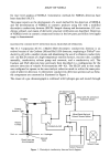
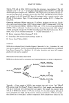
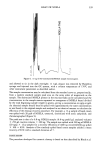
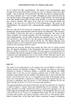
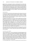
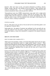
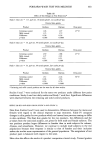
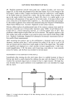
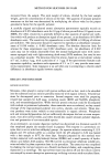
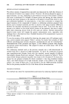
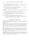
348 JOURNAL OF THE SOCIETY OF COSMETIC CHEMISTS vitiligo to normal skin, that there was significant protection afforded at 254 and 366 nm, while at 300 nm there was no significant protection. He further reported similar results with delayed pigmentation in normal volunteers. The aims of this study were (a) to determine if a correlation exists between melanin level in the skin and minimum erythema dose and (b) to determine the absorption of human melanin in the ultraviolet and thus arrive at an estimate of the photoprotection that it offers. MATERIALS AND METHODS MINIMUM ERYTHEMA DOSE DETERMINATION The minimum erythema dose was determined on each of 47 psoriatic patients about to start treatment with UVB. The phototests were conducted on skin sites that were not previously exposed to ultraviolet radiation. The irradiation was conducted using a bank of Philips TL12 4-foot fluorescent lamps, and the doses were from 10 to 150 mJ/cm 2 in steps of 10 mJ/cm 2. Each dose was delivered to an area of ! cm 2. An arithmetic series was chosen rather than a geometric series to avoid errors in irradiation times. The irradiance was measured with an International Light model 442 phototherapy radiom- eter with a model SEE 1240 detector. The skin reaction was assessed 24 hours later, and if no erythema was perceptible, the test was repeated in another area, preferably of the back, from !40 to 290 mJ/cm 2 in steps of 10 mJ/cm 2 and the reaction was assessed 24 hours later. This procedure was repeated until an erythema reaction was observed. This procedure was adopted as we found that it was difficult to predict with reasonable accuracy the MED by visual observation of the pigment level of the skin. At the time that the erythema reaction was assessed, a measurement was carried out to determine the pigment level in the skin adjacent to the irradiated area on a previously unexposed site (7). In some of the darker patients we had to determine the minimum perceptible erythema instrumentally, as it was difficult to discern it visually (8). MELANIN SPECTRUM DETERMINATION The apparent absorbance of human skin was determined by recording the diffuse reflec- tance from 275 to 720 nm (8), which was referenced to that of BaSO 4. The instrument used (Figure !) consisted of a !000-watt Xenon arc lamp with a light feedback stabi- lizer. The output of the lamp was focused through a 0.08-m water filter on the input Xe-Lamp ...m ....... •. Pr.o6• S•:mple Fief. Figure 1. A schematic diagram of the diffuse reflectance spectrophotometer.
Purchased for the exclusive use of nofirst nolast (unknown)
From: SCC Media Library & Resource Center (library.scconline.org)